

ULTIMA / Science & evidence
Natural expression.
A scientific
perspective.
Explore the science behind facial muscle conditioning, light-based skin care and the ULTIMA treatment experience. Original observations. Objective imaging. Research you can trace.
Ultima FACE - Professional Facial Muscle stimulation Technology
ULTIMA course evaluation14Selected labelled ultrasound
measurement sequences6Unique five-session
LifeViz 3D cases18Cases in the photographic
outcomes portfolio
Explore every chapter
Direct access to every topic and every source.Full document index — all 34 folder files + original evaluation
- S01 · Resource navigator
- S02 · ULTIMA educational presentation
- S03 · Theoretical training video
- S04 · IonLift treatment demonstration
- S05 · Professional sales presentation
- S06 · Educational presentation · PDF edition
- S07 · CELONIQUE product catalogue
- S08 · CELONIQUE formulation sheets
- S09 · CELONIQUE research protocol
- S10 · CELONIQUE anamnesis & evaluation forms
- S11 · Integrated scientific evidence report
- S12 · Dermus objective imaging report
- S13 · Facial NMES integrative review
- S14 · Photographic outcomes review
- S15 · Potential effects on topical delivery
- S16 · LifeViz facial 3D evaluation
- S17 · MuscleGuard concept presentation
- S18 · Face treatment results portfolio
- S19 · ULTIMA Shape treatment protocol
- S20 · Updated ULTIMA Face protocol
- S21 · Client procedure information
- S22 · Face & Shape preparation checklist
- S23 · IonLift user guide · English PDF
- S24 · IonLift user guide · editable English
- S25 · Potential effects on topical delivery · second copy
- S26 · IonLift user guide · Hungarian
- S27 · HILEFT & injectable treatment concept
- S28 · UL-800 operator manual · English
- S29 · UL-800 operator manual · Hungarian
- S30 · Client declaration · Hungarian DOCX
- S31 · Client declaration · short English version
- S32 · Client declaration · Hungarian PDF
- S33 · Client declaration · English DOCX
- S34 · Client declaration · English PDF
- S35 · Original 25-person evaluation
01 / The science of ULTIMA
An evidence-led approach
to a refreshed appearance.
ULTIMA combines professional facial muscle stimulation with an optical skincare concept. The evidence is easiest to understand through three complementary perspectives.
01 / Direct observations
What was recorded with ULTIMA?
Professional course evaluations and selected case series describe improvements in contour, firmness and a refreshed appearance. The results below identify the treatment, measurement method and source.
Discover the product-specific findings ↗02 / Objective imaging
Looking beyond the photograph.
Dermus ultrasound records local skin-thickness measurements. LifeViz 3D maps changes in facial surface shape. Together, they add distinct measurement perspectives to the aesthetic story.
Explore the imaging ↗03 / Published research
A technology with scientific context.
Peer-reviewed work on facial NMES, photobiomodulation and iontophoresis explains why these technologies are relevant. Each reference states how closely its device and protocol relate to ULTIMA.
Read the research connections ↗The four ULTIMA datasets describe different methods and may overlap in participants or source material. Their counts are kept separate. The supplied product-specific studies are professional evaluations and exploratory observations; published trials of other devices provide supporting context.
02 / The science of ULTIMA
Condition the muscles.
Support natural-looking contours.
The facial muscles contribute to expression and surface shape. Controlled electrical activation gives ULTIMA its distinctive muscle-focused aesthetic approach.
The rationale for facial EMS
Neuromuscular electrical stimulation applies pulses through skin-contact electrodes to produce controlled muscle activation. Repeated activation provides a rationale for facial muscle conditioning and the appearance of improved support and definition.
ULTIMA’s documented architecture uses a symmetric biphasic rectangular waveform, with selectable program parameters and sequential channel distribution. The protocol therefore describes a specific physical stimulus, rather than an unspecified “facial workout”.
The published facial NMES literature includes ultrasound and CT observations of muscle-related changes. These studies make the muscle-conditioning concept scientifically relevant; ULTIMA’s own appearance and imaging observations show how its treatment experience is being evaluated.
03 / The science of ULTIMA
Light-based care.
A deeper scientific context.
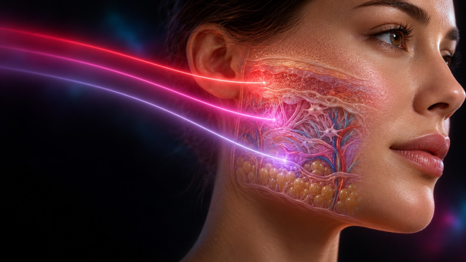
Red and near-infrared photobiomodulation research explores how light exposure can influence skin biology and appearance. ULTIMA’s optical concept draws on this field.
Why wavelength and dose matter
Photobiomodulation studies describe interactions between absorbed light and cellular signalling. In aesthetic research, repeated exposure has been evaluated using wrinkle measurements, skin-surface profiling, photography and ultrasound-related readouts.
The ULTIMA technical specification and integrated evidence report identify a 660 / 730 / 940 nm optical architecture. The 660 nm component has a direct wavelength connection to a randomized periocular-wrinkle study. Published red/near-infrared studies using other wavelengths add broader biological context.
For a meaningful comparison, wavelength must be considered alongside irradiance at the skin, exposure time, treatment area, duty cycle and the number of sessions. A shared wavelength is a relevant scientific connection; it does not by itself establish the same delivered dose or outcome.
04 / The science of ULTIMA
A considered next step
for professional skincare.
IonLift adds a dedicated topical-application stage to the ULTIMA experience, pairing the treatment workflow with selected CELONIQUE formulations.
The dedicated IonLift workflow
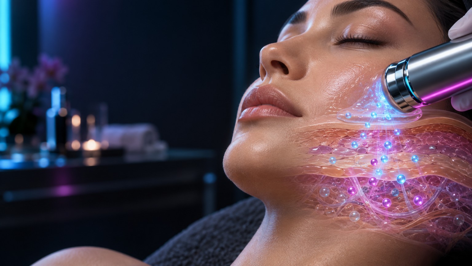
The guide describes a ten-minute adjunct after the facial session, using one or two moving applicators and a handheld return electrode. It specifies dedicated IonLift programs, product-assisted skin contact and a controlled warming function.
Iontophoresis research offers a scientific basis for investigating electrically assisted transport of selected ingredients. The behaviour of any formulation depends on its molecular properties and on the actual electrical delivery conditions.
The CELONIQUE sheets identify humectants, niacinamide, hyaluronic-acid forms, peptides and plant-derived vesicle ingredients. They connect the skincare concept to defined formulations. Direct uptake and depth measurements would be needed to quantify the delivery of particular ingredients during this combination treatment.
05 / The science of ULTIMA
A standardized course.
A visible aesthetic signal.
The original professional evaluation follows twenty-five adults through a defined Contour Lift course, combining before/after photography with participant feedback.
ULTIMA-specific · Single-arm evaluation
At least noticeable appearance improvement
The report describes approximately 68% with clearly visible contour improvement and 20% with mild but noticeable improvement after the course.
Percentages are reproduced as approximate values from the source. This was a manufacturer-supplied professional evaluation without an untreated or sham comparator.
A clearly described treatment routine
Twenty-five adults completed the course. Twelve participants under 45 received five sessions; thirteen aged 45 or over received seven sessions. Each session used Contour Lift for 25 minutes, around twice weekly, with at least 72 hours between visits.
The report describes consistent photography, neutral expression and comparison of contour, firmness and overall refreshed appearance. All participants reported treatment value: approximately 80% found the course satisfactory and 20% preferred additional sessions.
Two participants reported mild initial pinching or stinging that diminished during the session. One temporary local skin response resolved in about 36 hours. No serious adverse events were reported in this small evaluation.
06 / The science of ULTIMA
Objective imaging.
The immediate skin response.
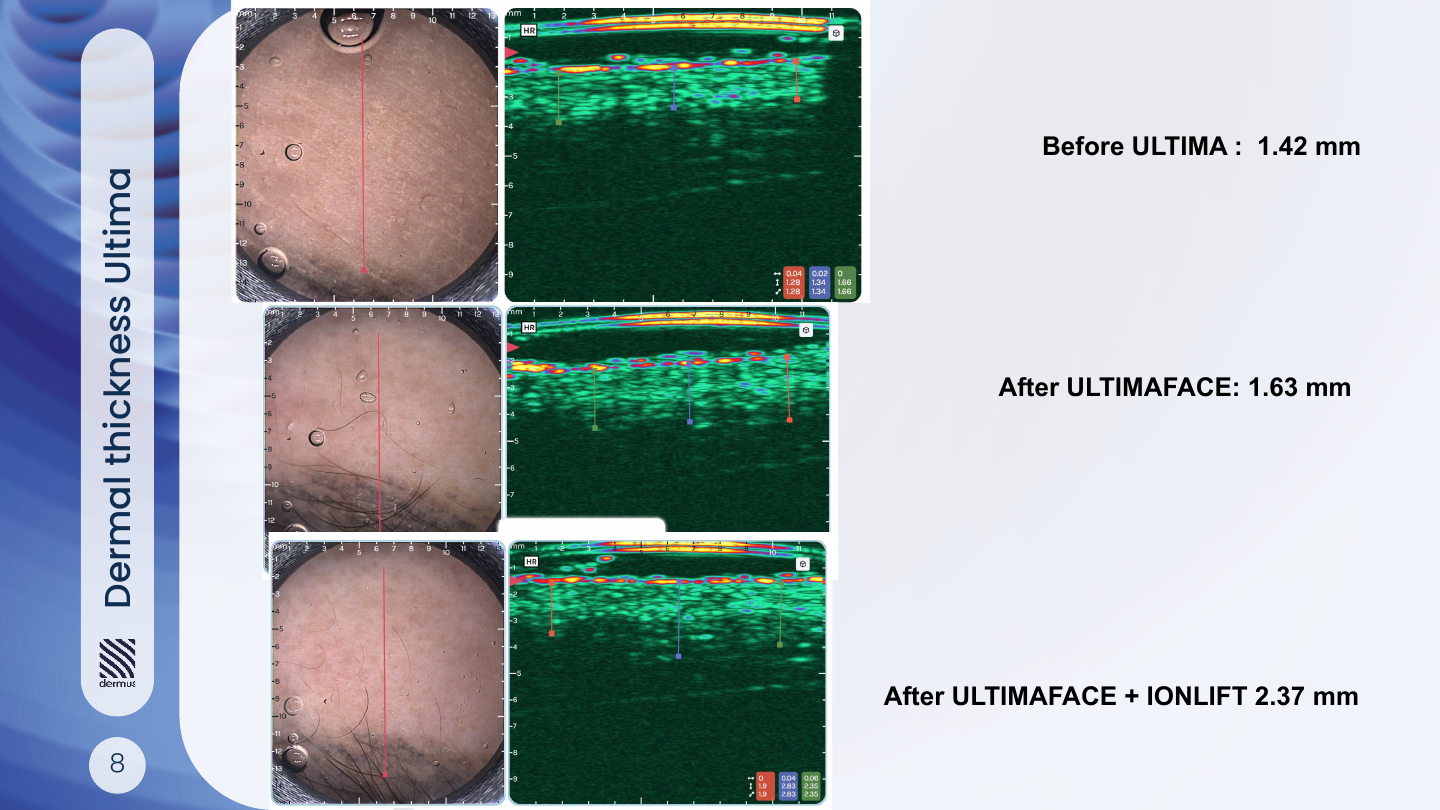
Dermoscopy-guided ultrasound adds a subsurface measurement to the ULTIMA evidence portfolio. The report documents local changes in measured dermal thickness.
Fourteen labelled comparisons
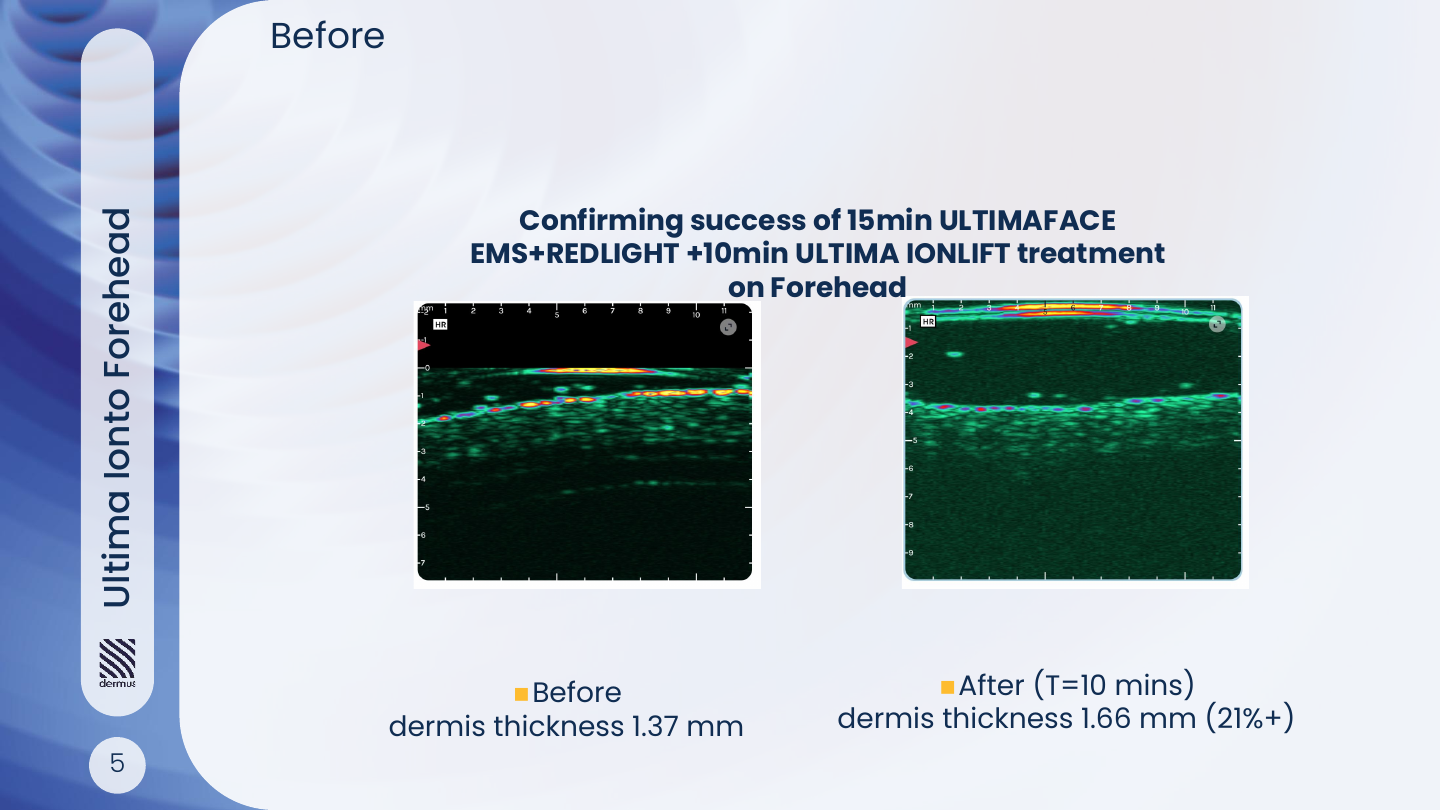
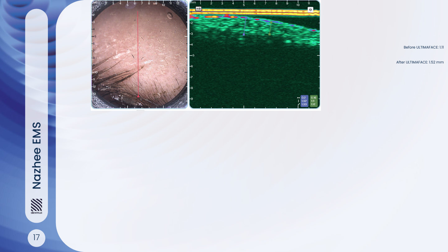
The report consolidates fourteen selected baseline-to-post sequences recorded with Dermus high-frequency ultrasound. The principal workflow involved a 15-minute ULTIMAFACE step, with an optional ten-minute IonLift stage. Measurements are reported in millimetres.
The selected comparisons show a positive thickness change, with a reported median of +15.2%. The report’s unit of analysis is a measurement sequence: fourteen sequences must not be presented as fourteen independent participants or a universal treatment success rate.
The immediate timing is compatible with hydration, fluid redistribution and other acute local responses. Establishing longer-term tissue remodelling requires repeat follow-up and suitable controlled measurements.
Open all 14 measurements and calculation notes
| Source label | Reported treatment context | Baseline, mm | Post, mm | Change |
|---|---|---|---|---|
| S05 | Forehead · ULTIMAFACE + IonLift | 1.37 | 1.66 | +21.2% |
| S06 | Cheek · ULTIMAFACE + IonLift | 2.71 | 2.86 | +5.5% |
| S07 | Cheek · ULTIMAFACE + IonLift | 1.27 | 1.60 | +26.0% |
| S08 | Sequential EMS + IonLift | 1.42 | 2.37 | +66.9% |
| S09 | IonLift | 1.71 | 1.96 | +14.6% |
| S10 | Sequential treatment | 1.43 | 1.54 | +7.7% |
| S11 | Zygomatic · EMS | 1.58 | 1.64 | +3.8% |
| S15 | Combined treatment | 1.96 | 2.27 | +15.8% |
| S16 | EMS | 1.19 | 1.50 | +26.1% |
| S17 | EMS | 1.11 | 1.52 | +36.9% |
| S18 | Combined treatment | 2.26 | 2.75 | +21.7% |
| S19 | Cheek · combined | 1.69 | 1.76 | +4.1% |
| S20 | Forehead · EMS | 1.62 | 1.84 | +13.6% |
| S21 | IonLift · supplementary timepoint | 2.15 | 2.44 | +13.5% |
The reported median of 15.2% is the median of the fourteen tabulated percentage changes. S08 uses the final combined reading. S21 is a supplementary timepoint: the source table and prose describe its timing differently, so no exact duration is assigned here. These selected sequences are not pooled into a participant-level efficacy estimate.
07 / The science of ULTIMA
Mapping change
in three dimensions.
Six LifeViz cases extend the evidence from a flat photograph to regional facial surface comparisons after five ULTIMAFACE sessions.
The clearest pattern is in the midface
The review identifies localized positive surface displacement around the cheeks in several cases. F-03 has the most balanced bilateral pattern, while F-02, F-04 and F-06 provide additional cheek-region examples.
In F-03, the selected cheek regions carry displayed values of 0.83 and 0.94. The figure labels these regional values in mL and also displays a separate colour scale in millimetres. The report asks for confirmation of the underlying export and ROI definitions; these values are therefore retained as software annotations, not interpreted as newly formed tissue.
Expression-sensitive regions, including the lips, eyelids and brows, need particularly consistent positioning. The six-case material supports further study of contour change, with standardized capture and repeatability testing as the next step.
| Case | Selected observation | Interpretive context |
|---|---|---|
| F-01 | Prominent perioral comparison | Lip posture makes this region expression-sensitive. |
| F-02 | Localized cheek change | Supportive of the midface pattern. |
| F-03 | Bilateral midcheek comparison | Strongest balanced example in the review. |
| F-04 | Lateral cheek comparison | Regional finding; capture alignment matters. |
| F-05 | Forehead and perioral readings | Brow and lip position affect interpretation. |
| F-06 | Bilateral cheek comparison | Additional midface example after five sessions. |
08 / The science of ULTIMA
A consistent appearance story
across selected cases.
The photographic portfolio brings the evidence back to the client experience: contour, a firmer-looking midface and a refreshed expression.
Eighteen cases, individually described
The supplied review covers clients aged 35–76, with a mean age of 50.6 and a median of six sessions. Twelve clients received five or six treatments, five cases show a single-session comparison, and one case documents thirty sessions.
Lower-face definition and midface appearance recur across the source descriptions. The single-session examples illustrate an immediate refreshed appearance; the repeated-course examples provide a broader view of the treatment experience.
The assessment is descriptive and based on selected images, with lighting, head position and expression influencing comparison. The source reports positive client feedback, but does not provide a blinded controlled evaluation.
Read all 18 coded case summaries
| Case | Age | Sessions | Appearance observations reported in the source |
|---|---|---|---|
| P01 | 54 | 6 | Jawline and submental definition; fuller-looking midface |
| P02 | 65 | 6 | Softer nasolabial appearance; brow and midface refresh |
| P03 | 51 | 5 | Facial contour and cheek appearance |
| P04 | 43 | 1 | Immediate nasolabial and upper-face refresh |
| P05 | 56 | 6 | Firmer-looking skin and midface |
| P06 | 59 | 5 | Brow, midface and jawline definition |
| P07 | 35 | 1 | Immediate refreshed eye area and contour |
| P08 | 57 | 1 | Forehead, under-eye and jawline appearance |
| P09 | 38 | 1 | Immediate jawline and lower-face definition |
| P10 | 39 | 6 | Fine-line and midface appearance |
| P11 | 38 | 6 | Jawline, brow and upper-eyelid appearance |
| P12 | 76 | 6 | Brow, midface and firmness |
| P13 | 49 | 6 | Periocular and upper-eyelid appearance |
| P14 | 40 | 6 | Periocular, jawline and upper-lip contour |
| P15 | 62 | 6 | Periocular and nasolabial appearance |
| P16 | 38 | 5 | Under-eye, midface and jawline appearance |
| P17 | 61 | 30 | Facial balance and eye-area appearance |
| P18 | 49 | 1 | Immediate jawline and submental definition |
09 / The science of ULTIMA
Building the next chapter
with CELONIQUE.
The research programme connects facial stimulation, professional skincare and structured assessment in a developing combination-treatment project.
Shanghai: a documented research setting
ULTIMA’s public update of 16 February 2026 identifies MAIO Beauty Clinic in Shanghai, with the professional support of Royal Life Group, as the setting for the ongoing CELONIQUE collaboration. It reports forty participants and favourable interim observations of appearance and treatment experience.
The supplied October 2025 protocol describes an earlier planned sixty-participant, four-group pilot across Germany and China. That planned design and the later forty-participant public update are presented as separate milestones. The reviewed folder contains the protocol and collection forms, rather than a completed comparative results dataset.
The formulation documents make the skincare component identifiable. Together with the ultrasound examples, the project provides a useful platform for studying how the combined professional workflow performs over repeated visits.
10 / The science of ULTIMA
Make the scientific
connection precise.
A convincing technology comparison identifies what is shared, what differs and which conclusions can be carried across.
Documented ULTIMA facial programs
| Program · 25 minutes | Frequency | Pulse width | Work / pause | Ramp up / down | Source page |
|---|---|---|---|---|---|
| Contour Lift 25 | 112 Hz | 280 µs | 8 / 0.5 s | 1.2 / 0.8 s | p. 7 |
| Rebuild Tone 25 | 76 Hz | 360 µs | 8 / 0.5 s | 1.8 / 0.8 s | p. 8 |
| Face Sculpt 25 | 220 Hz | 200 µs | 12 / 0.5 s | 1.2 / 0.6 s | p. 15 |
| Power Contour 25 | 180 Hz | 480 µs | 6 / 0.5 s | 1.8 / 0.4 s | p. 16 |
How the external research relates
| Research connection | External study / delivery | ULTIMA documentation | What the comparison supports |
|---|---|---|---|
| Facial NMES · R01 | 20 min, 5 days/week, 12 weeks; zygomatic endpoint | Contour Lift: 112 Hz, 280 µs, 25 min; professional course | Shared muscle-activation principle; device and cumulative exposure differ. |
| Periorbital NMES · R02 | Thin-film electrode; 10 min, 3 times/week, 8 weeks | Multi-region facial applicator and different program family | Anatomical rationale for muscle-associated contour change. |
| High-frequency fNMES · R03 | CERTEC: 40–190 kHz | UL-800 specification: 4–220 Hz | Different operating regimes; no matched waveform or output claim. |
| 660 nm PBM · R06 | 660 nm; about 3.8 J/cm²; 10 sessions / 4 weeks | 660 / 730 / 940 nm listed in technical documentation | Direct red-wavelength overlap; irradiance, dose and geometry require separate matching. |
| Red/NIR combination · R08 | 633 / 830 nm; 126 / 66 J/cm² | 660 / 730 / 940 nm architecture | Related optical concept; wavelengths and exposure protocol are not identical. |
| Iontophoresis · R10 | Transport depends on current, polarity and formulation | Dedicated IonLift step, moving heads and return electrode | Relevant transport mechanism; no assumption of identical molecule delivery. |
For an optical exposure, energy density (J/cm²) equals irradiance (W/cm²) multiplied by time (s), with duty cycle considered where relevant. A complete comparison also records measurement distance, spectral contribution, applicator area and output calibration.
11 / The science of ULTIMA
A shared platform.
A distinct body-treatment programme.
ULTIMA Shape extends the professional platform to body-focused muscle conditioning, with named programs and original IR1–IR4 applicator configurations.
The supplied Shape guide defines six 25-minute programs with different pulse widths, frequencies and contraction patterns. This provides a reproducible technical framework for targeted conditioning and future objective evaluation.
The body protocol is included here as part of the complete knowledge base. Face-study outcomes remain facial evidence; a body-specific outcome claim requires a body-specific dataset. The strongest current contribution of this source is its precise description of how the intended treatment stimulus is delivered.
| Program | Frequency | Pulse width | Applicator context in the guide |
|---|---|---|---|
| Power Sculpt 25 | 92 Hz | 420 µs | IR3 / IR4 · larger muscle areas |
| Flat Core 25 | 92 Hz | 360 µs | IR1, or IR1 + IR2 · abdomen |
| Glute Lift 25 | 92 Hz | 450 µs | IR1 + IR2 · gluteal placement |
| Shape Duo 25 | 84 Hz | 360 µs | Combined IR1–IR4 configurations |
| Firm & Shape 25 | 36 Hz | 250 µs | IR1 + IR2 · specified placement |
| Deep Recovery 25 | 16 Hz | 180 µs | IR3 / IR4 · gluteal placement |
12 / The science of ULTIMA
Professional care
is part of the evidence story.
Clear screening, consistent preparation and client feedback make treatment delivery more reproducible and the experience easier to evaluate.
Prepare
Start with the individual.
The supplied declarations and checklist support a consultation, suitability screening, clean skin, correct coupling and the relevant eye-protection procedure. Current approved instructions guide the trained operator’s decisions.
Personalize
Monitor comfort throughout.
Program selection, contact and stimulation intensity are adjusted within the appropriate instructions. The client-information sheet emphasizes communication and a comfortable experience, with prompt adjustment if discomfort occurs.
Document
Build a meaningful record.
Consistent photographs, session details, feedback and follow-up timing make results easier to compare. Reusable applicators also have defined cleaning, drying and inspection procedures in the operator documentation.
13 / The science of ULTIMA
Read the research.
Understand the relevance.
Each reference explains its design, result and connection to ULTIMA. Studies of other systems are clearly identified as supporting technology or measurement evidence.
Facial muscle toning: randomized controlled trial
In a randomized, partially blinded study of 108 women, twelve weeks of facial NMES was associated with an 18.6% increase in zygomatic-major thickness in the treatment group. Treatment lasted 20 minutes a day, five days per week. The study supports the principle that repeated electrical activation can condition facial muscle, which is central to ULTIMA’s approach. The device, exposure schedule and electrode arrangement differ, so 18.6% is the result of that trial and is not an ULTIMA outcome.
Objective CT analysis of facial muscle morphology
This prospective split-face pilot analysed fourteen women after eight weeks of treatment with a periorbital thin-film NMES device. CT measurements found increased orbicularis-oculi thickness and improvements in the corresponding contour measurements on the treated side. The study strengthens the anatomical rationale for considering muscles as part of facial rejuvenation. Its 10-minute sessions three times weekly, targeted electrodes and device differ from ULTIMA, so the connection is mechanistic rather than proof of equivalent performance.
Facial wrinkles and sagging: split-face study
The eight-week study involved twenty-four women and reported improvements in facial elasticity, wrinkle measures and contour-related outcomes. It used a CERTEC device with a 40–190 kHz operating range, which is materially different from ULTIMA’s documented Hz-range programs. It broadens the evidence that electrical approaches can influence facial appearance, while supporting no direct parameter equivalence. The March 2026 correction addresses conflict-of-interest disclosure and the ethics approval number; both records should be read together.
Focused electric field technology and muscle rejuvenation
A randomized, self-controlled clinical experiment in thirty-one women examined a wearable focused-electric-field device alongside separate laboratory and animal work. The human study reported improvements in skin elasticity and selected lifting-related measures. It offers relevant context for ULTIMA’s muscle-focused aesthetic concept. The field delivery and device differ, and the laboratory mechanisms do not establish satellite-cell activation or muscle regeneration in ULTIMA clients.
Neuromuscular electrical stimulation in facial aging
This integrative review links facial anatomy with the rationale for neuromuscular stimulation and identified two eligible clinical studies. It includes positive zygomatic-muscle findings alongside a smaller study with different masseter results. For ULTIMA, the review supports the relevance of muscle-specific protocols and objective follow-up. Its value is a scientific framework for interpreting facial EMS, with the clinical evidence still dependent on the particular device and study design.
Red and amber light for periocular wrinkles
This randomized split-face study recruited 137 women and compared 660 nm red with 590 nm amber light over ten sessions in four weeks at approximately 3.8 J/cm². The reported wrinkle-volume reductions were 31.6% and 29.9%, respectively; hydration and viscoelasticity did not improve. The 660 nm arm provides a direct wavelength-level connection to the three-wavelength ULTIMA specification. Dose, geometry and protocol are different, so the study supports the red-light rationale without making those percentages ULTIMA results.
Controlled red and near-infrared photorejuvenation trial
This controlled study enrolled 136 volunteers and evaluated 30 sessions of 611–650 nm or 570–850 nm light against controls. Photography, profilometry and ultrasound-based measures documented improvements in skin appearance, roughness and collagen-density-related readings. It supports a biological and measurement framework for ULTIMA’s light component. Its broad spectra, treatment geometry and repeated exposure differ from ULTIMA; the longer-term outcomes also cannot be inferred from an immediate Dermus thickness change.
Combined 633 nm and 830 nm LED treatment
Thirty-one participants received nine LED treatments combining 633 nm and 830 nm exposures, with follow-up skin-profile and photographic assessments. The study reported improved photoaging measures and favourable participant assessments. It supports the concept of a red/near-infrared skincare approach relevant to ULTIMA’s optical rationale. The wavelengths, 126/66 J/cm² exposures and Omnilux hardware are different, so this is supportive technology evidence rather than a matched ULTIMA protocol.
Light parameters and photobiomodulation efficacy
This review examines why photobiomodulation depends on several interacting exposure parameters. It provides a useful framework for comparing wavelength, irradiance, energy density, pulse characteristics and treatment timing. For ULTIMA, it supports documenting the actual applicator and selected light settings alongside outcomes. Matching one wavelength establishes a relevant connection, while a reliable efficacy bridge also requires compatible dose and delivery conditions.
Iontophoresis-assisted transdermal delivery
The review describes how low-level electrical currents can assist the movement of selected substances across the skin barrier. It provides mechanistic context for the dedicated IonLift concept and highlights the importance of formulation and electrical delivery. Its main focus is drug-delivery research in inflammatory skin conditions, rather than cosmetic ULTIMA outcomes. It does not demonstrate that the particular CELONIQUE vesicles reach a specified depth during an ULTIMA session.
High-frequency ultrasound for evaluating skin aging
This review discusses high-frequency ultrasound as a way of characterizing skin aging and evaluating aesthetic products and procedures. It supports the choice of an objective imaging method for longitudinal skin assessment. For ULTIMA, the connection is to the measurement strategy used in the Dermus project. It validates the relevance of the method, rather than proving the efficacy of the treatment being measured.
Ultrasound in Aesthetics: Filler and Non-Filler Applications
This publication surveys aesthetic applications of ultrasound, including uses beyond fillers. It provides context for imaging skin and superficial structures in professional aesthetic work. Its relevance to ULTIMA is methodological: imaging can document changes that ordinary photographs do not measure. Treatment effectiveness still depends on the quality, reproducibility and design of the ULTIMA-specific evaluation.
LifeViz 3D imaging and standardized photography
LifeViz is a 3D photography platform for evaluating facial surface appearance and volume-related changes. Its documentation explains why positioning and consistent capture are important for repeat comparisons. This is directly relevant to interpreting the six ULTIMA LifeViz cases and their regional comparison maps. The imaging-system manufacturer’s information supports the measurement method and is not an independent endorsement of ULTIMA efficacy.
Additional background links retained from the educational deck
The August 2025 training deck also links to broader photobiology, wound-healing, exercise and connective-tissue literature. These links are preserved as background reading; they are not used to claim wound treatment, body-wide exercise effects or matched ULTIMA efficacy.
- Training slide 17 · additional photobiomodulation article (PMC5332991) ↗
- Chaves et al. · Light therapy and wound healing (2014) ↗
- Training slide 17 · DOI 10.1007/s10103-018-2572-z ↗
- Training slide 17 · DOI 10.1007/s10103-018-2526-5 ↗
- NIH · Understanding how exercise affects the body (2024) ↗
- Molecular mechanisms of stress-responsive collagen and elastin changes ↗
14 / The science of ULTIMA
The complete
source library.
Every file in the specified folder and its five subfolders is represented below, with an English summary, its evidence role and a direct source link.
15 / The science of ULTIMA
A clearer understanding.
Answers to the questions that matter when interpreting the science and discussing the treatment.
What scientific evidence is available specifically for ULTIMA?
The supplied material includes a 25-person standardized course evaluation, an eighteen-case photographic portfolio, six five-session LifeViz 3D cases and a selected fourteen-sequence Dermus ultrasound report. These are complementary professional and exploratory observations. Their design and findings are described separately above.
How do published studies of other devices support ULTIMA?
They establish relevant scientific context for facial electrical stimulation, photobiomodulation and iontophoresis. A useful bridge compares the physical delivery, anatomical target, dose and treatment course. The reference summaries identify those links and keep external study percentages attached to the devices actually tested.
Does increased ultrasound thickness mean new collagen formed immediately?
The Dermus measurements document an immediate local thickness response. Hydration, fluid distribution and perfusion-related changes are possible contributors. Establishing collagen remodelling requires appropriate longitudinal or tissue-specific endpoints.
What does the Shanghai study add?
It documents an active collaboration between ULTIMA, CELONIQUE and a Shanghai clinic, together with a protocol and participant-assessment framework. The February 2026 public update reports positive interim observations. A completed comparative dataset was not present in the reviewed folder.
How does this relate to a natural-looking lift or a red-carpet facial?
ULTIMA’s case descriptions emphasize a more defined-looking contour and a refreshed expression. A red-carpet facial is a service positioning around that experience. Individual response, the appropriate course and maintenance should be discussed with the treating professional.
Can I read the evidence page without internet?
Yes. All page text, menus, filters, fonts and displayed figures are included locally. Links to original Drive documents, journals and the ULTIMA website open external sources and require an internet connection.
Discover ULTIMA
with the evidence in view.
Explore a professional treatment approach centred on facial conditioning, natural-looking definition and a considered skincare experience.