

DOCUMENTED CASES
Before and after
See the change.
Understand its context.
Original photographs, LifeViz 3D comparisons and Dermus ultrasound images. Three perspectives that help you explore the changes associated with the ULTIMA treatments presented.
PERSPECTIVES IN THE GALLERY
18
Photographic cases
Comparing appearance.
04
LifeViz 3D panels
Spatial changes in the facial surface.
05
Dermus panels
Local ultrasound examination of the skin.
+ 4 featured case presentations with detailed explanations.
TAKE A CLOSER LOOK
Change becomes clear
in the details.
Choose an image type, then click an image to open the full panel. Images are presented in their original compositions; the arrangement of before-and-after views may differ between panels.
Photographic comparisons
Eighteen cases with different baseline conditions and treatment counts. Age and the number of treatments are stated for each image. The images present individual observations; they do not promise identical results for everyone.
ULTIMA FACE works through targeted, repeated activation of the facial muscles beneath the skin. Muscle conditioning aims to support the tissues and promote a firmer, more lifted appearance. The assessments below describe changes visible in photographs; muscle thickness, circulation and the extent of tissue elevation require separate measurements. Lighting, head position and facial expression also influence comparisons.
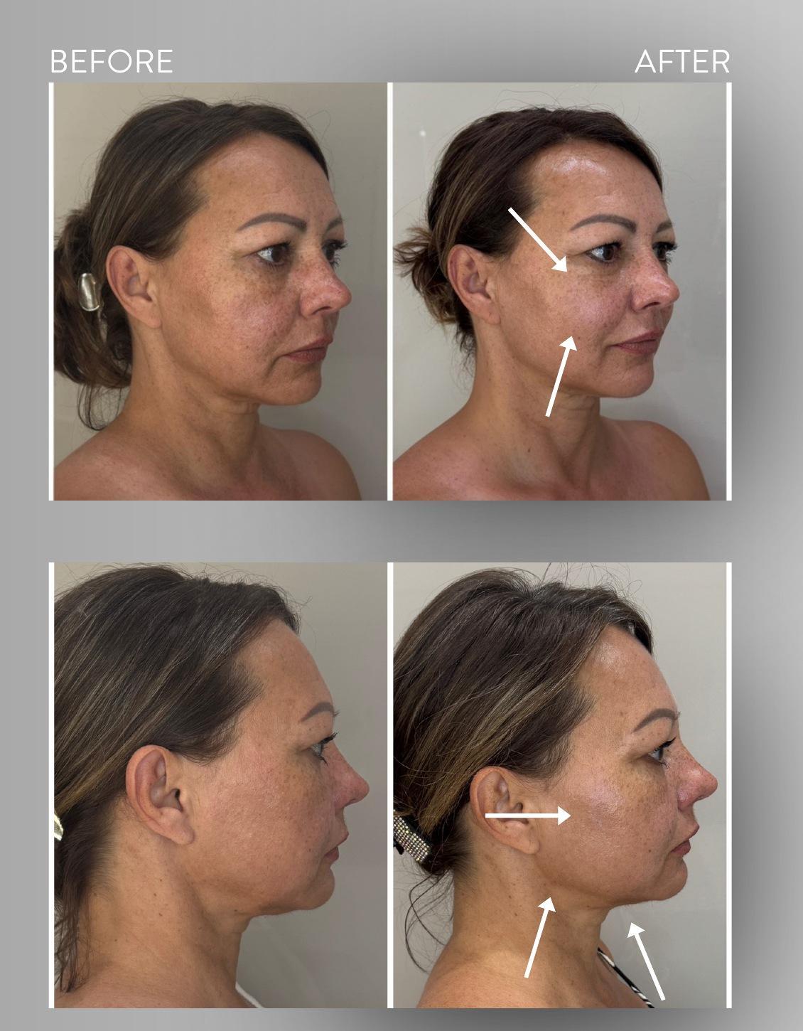
Case 01 · age 54 · 6 treatments
Lower face and profile
A more defined jawline and fuller-looking cheeks. After six sessions, both views show a clearer mandibular outline and under-chin transition. The midface appears fuller and more lifted, the cheek surface looks firmer, and the under-eye area appears fresher. This contour change aligns with the treatment goal of supporting the tissues through targeted facial muscle conditioning.
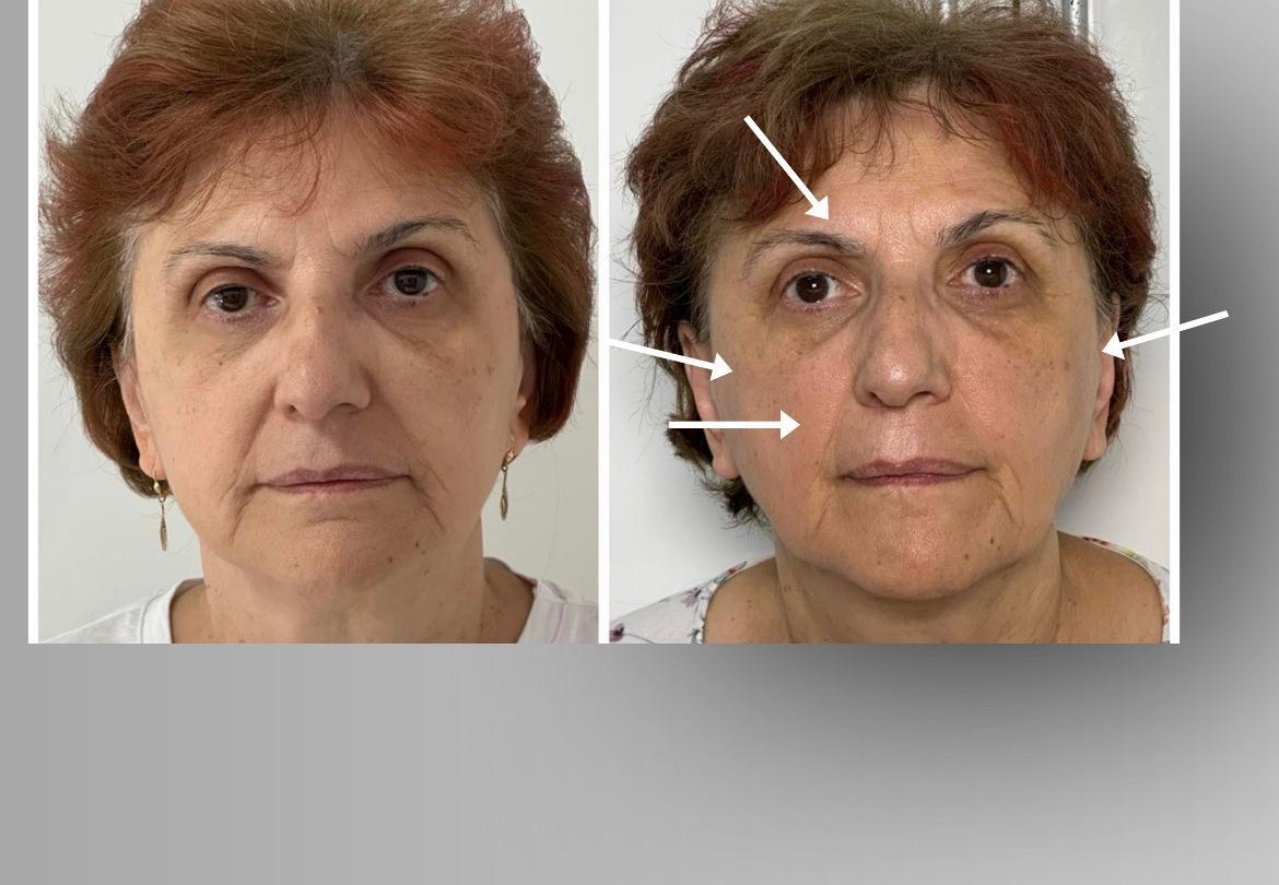
Case 02 · age 65 · 6 treatments
Midface and facial expression
Softer folds and a more harmonious midface. The six-session comparison shows less pronounced nasolabial folds, a smoother cheek transition and a more open-looking brow and eye area. The fuller, more lifted midface creates a more balanced appearance. These visible changes align with the aesthetic aim of treatment targeting the cheek muscles.
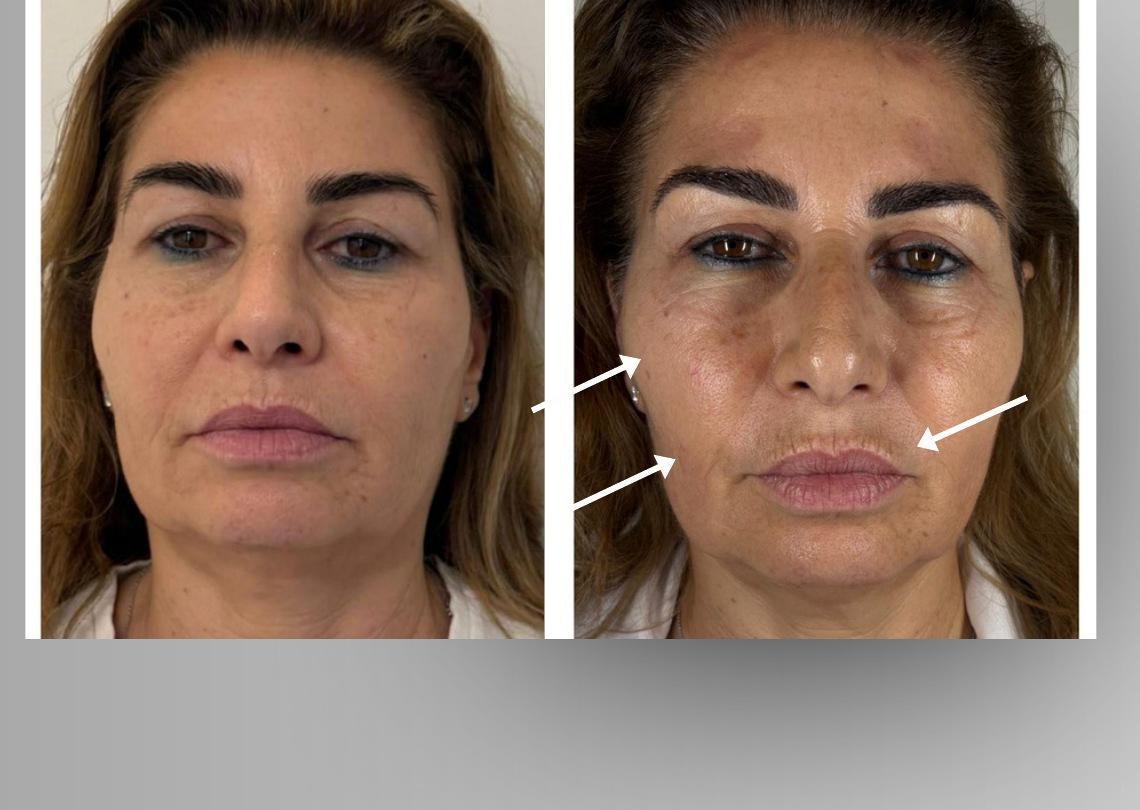
Case 03 · age 51 · 5 treatments
Facial contour
A more balanced contour and more prominent cheeks. After five sessions, the midface looks fuller and the cheek surface firmer; the lines around the mouth and the two sides of the face appear more balanced. A more lifted cheek contour gives the face a fresher appearance. This overall effect illustrates the contour-supporting aim of muscle conditioning.
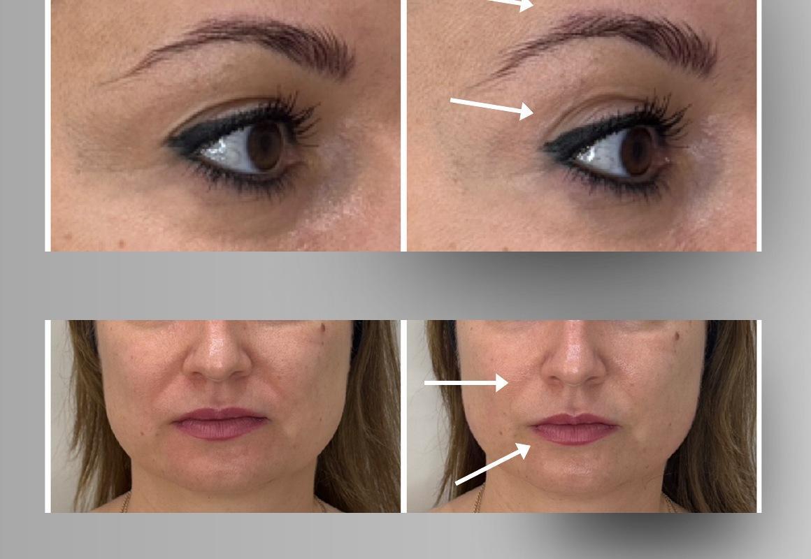
Case 04 · age 43 · 1 treatment
Change after one session
A more open-looking eye area after one session. In the close-up, the brows appear slightly higher and the eye area looks more open. The transition around the mouth looks smoother and the lower face more balanced. This is an immediate post-treatment appearance, consistent with a refreshing response to targeted muscle activation; the photographs do not measure lasting muscle remodelling.
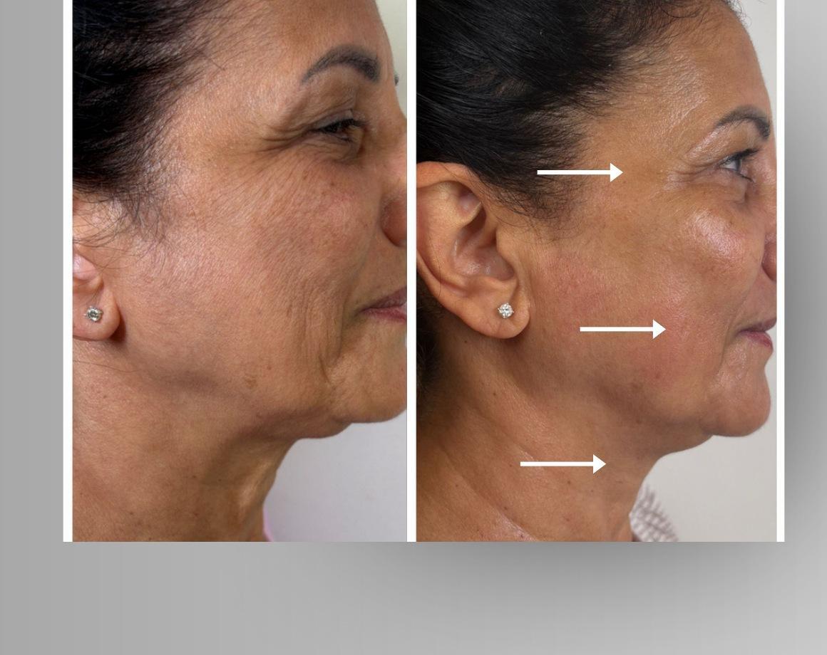
Case 05 · age 56 · 6 treatments
Profile and eye area
A smoother-looking eye area and firmer lower face. After six sessions, crow’s feet and fine surface lines on the cheek appear softer. The midface looks fuller and more lifted, with a more defined under-chin area. These combined surface and contour changes align with a muscle-conditioning approach; differences in expression also affect the eye-area comparison.
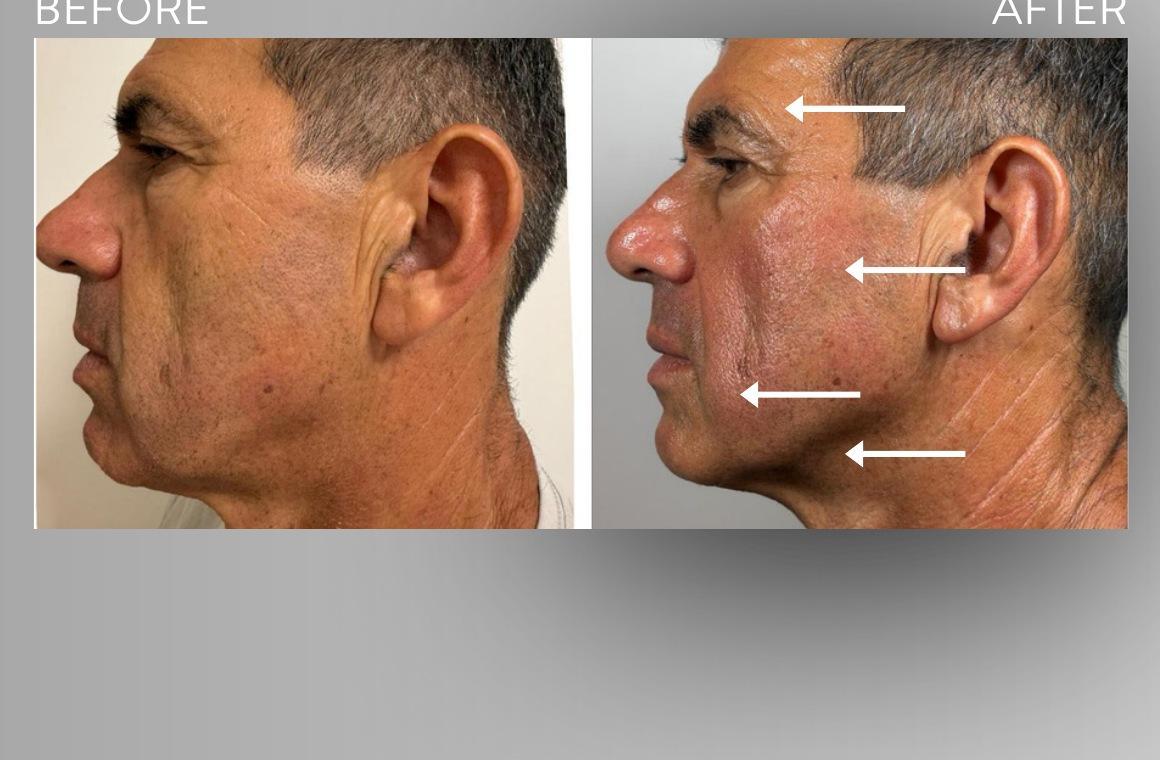
Case 06 · age 59 · 5 treatments
Male facial contour
A more defined masculine jaw contour. The profile after five sessions shows a sharper mandibular edge and under-chin boundary. The cheek appears better supported, the midface firmer and the lower face less heavy. The principal visible change is improved contour, which relates to the tissue-supporting aim of deeper facial muscle work.
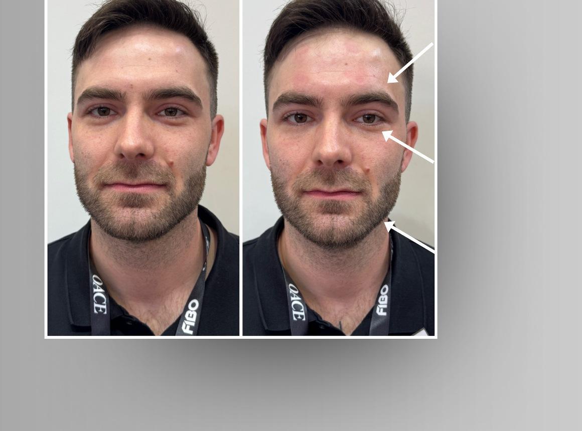
Case 07 · age 35 · 1 treatment
Single-session comparison
A fresher under-eye area and more refreshed appearance. After one session, under-eye shadow appears less pronounced and the brows slightly higher. The facial outline looks more defined. This illustrates immediate freshness following targeted muscle activation. The redness visible immediately after treatment is a temporary pressure mark left by the electrodes and fades shortly afterwards.
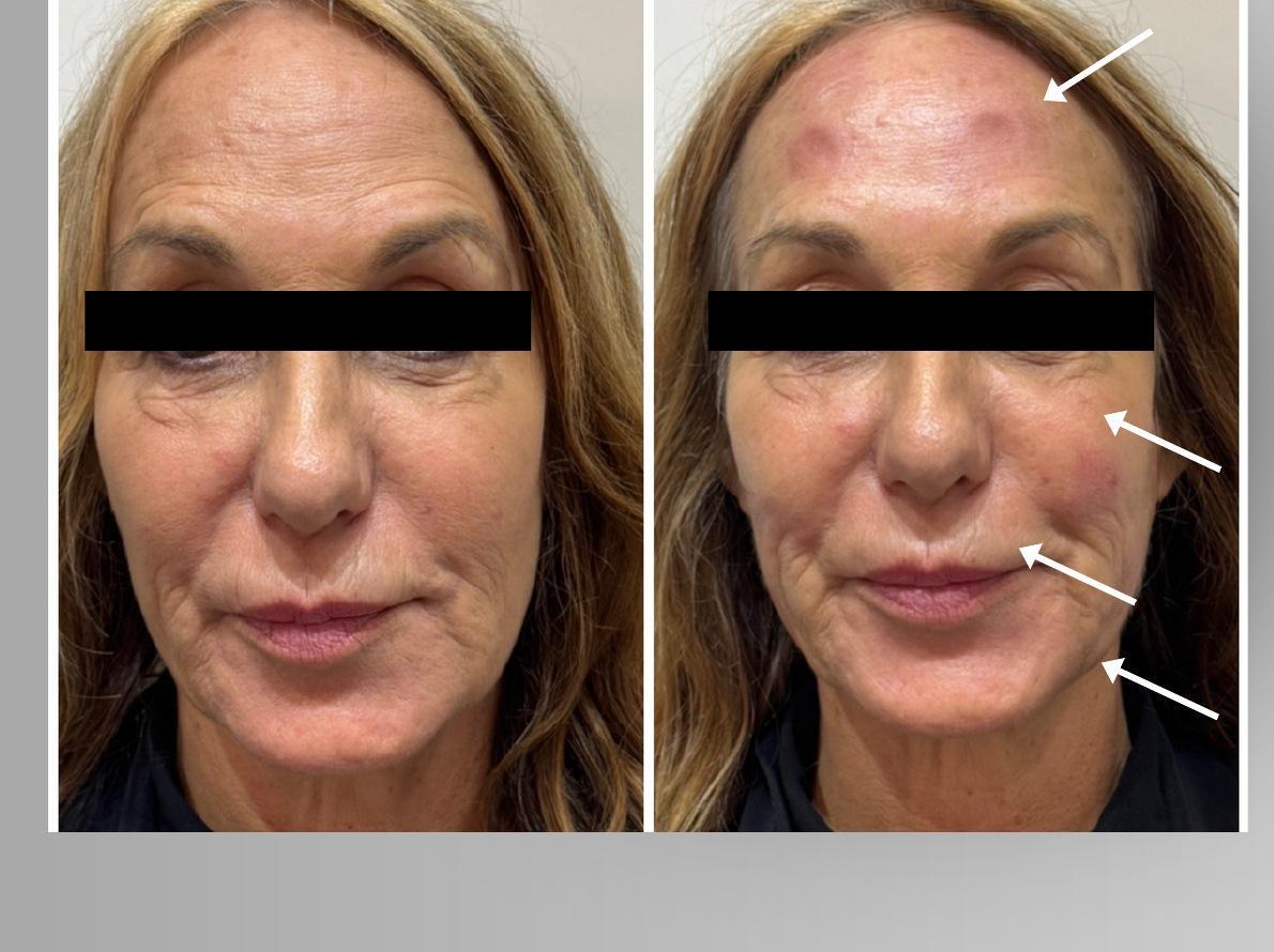
Case 08 · age 57 · 1 treatment
Single-session comparison
Softer forehead and upper-lip lines. After a single session, forehead folds and fine lines above the upper lip appear smoother, with a more refined jawline. This refreshed appearance documents the immediate response following facial muscle activation. The redness visible immediately after treatment is a temporary pressure mark left by the electrodes and fades shortly afterwards.
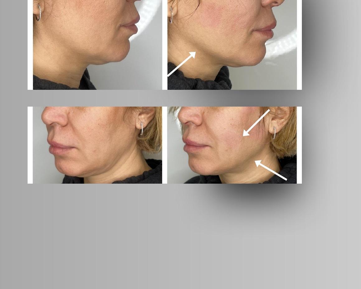
Case 09 · age 38 · 1 treatment
Profile comparison
A clearer lower-face contour from two angles. After one session, the jawline is more distinct from the under-chin area and the cheek-to-jaw transition looks firmer. The skin surface appears smoother and the contour more lifted. This is an immediate contour response following targeted muscle activation; the difference in head position must also be considered.
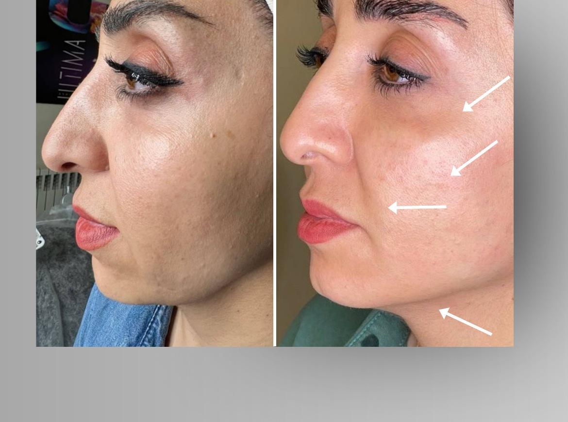
Case 10 · age 39 · 6 treatments
Comparison following a treatment course
A smoother cheek-to-fold transition and clearer jawline. After six sessions, the nasolabial shadow is softer, the cheek surface looks more even and the midface appears more lifted. The mandibular edge is more clearly defined. Changes across several facial areas align with the goal of repeated facial muscle work: supporting contour and the overlying tissues.
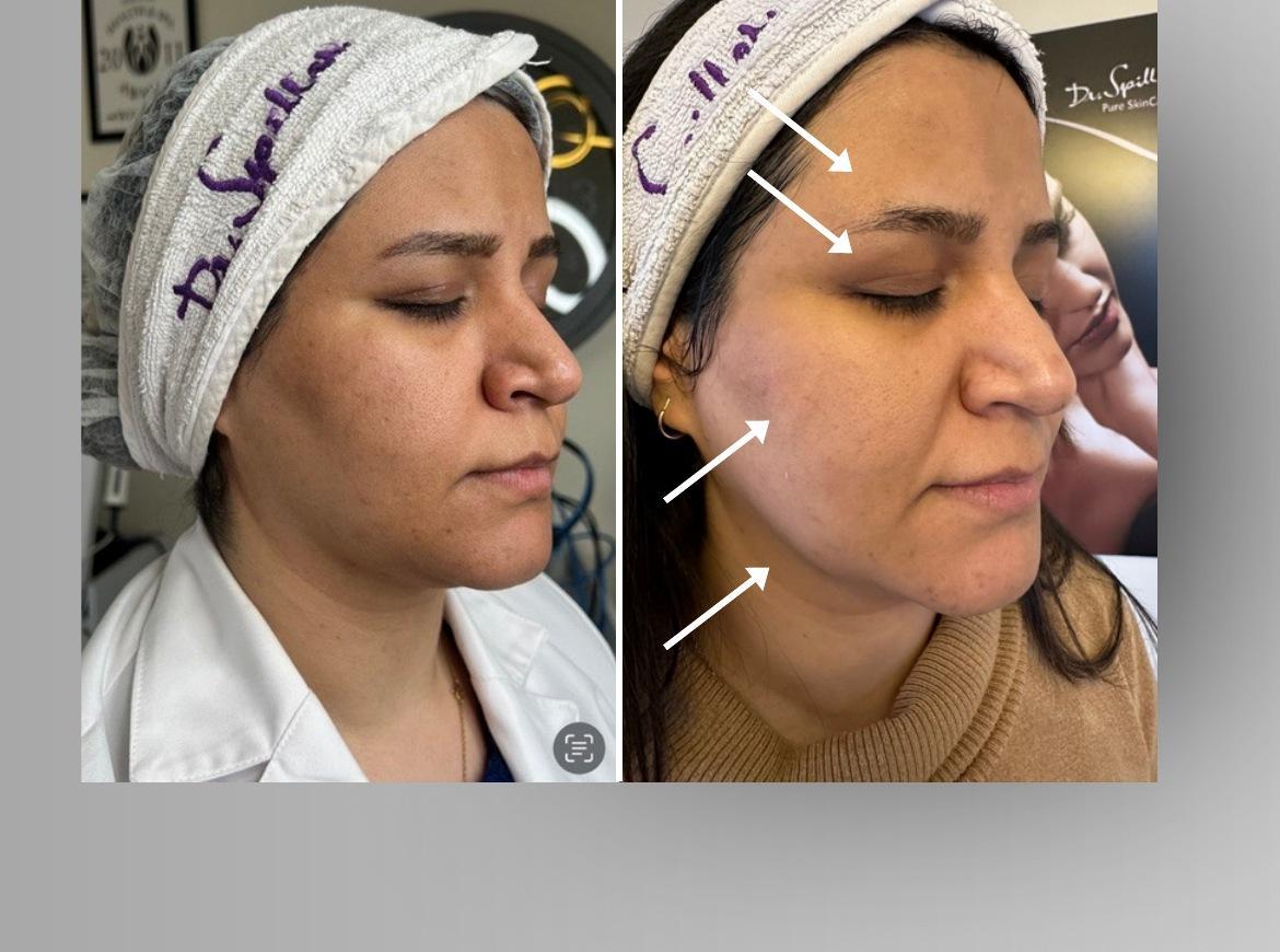
Case 11 · age 38 · 6 treatments
Facial appearance
A firmer cheek contour and more defined lower face. After six sessions, the cheeks look fuller, the jawline more defined and the skin surface smoother. The brow area appears more lifted; eye openness cannot be assessed because the eyes are closed. The main result is improved-looking contour and tissue support, consistent with the aim of muscle conditioning.
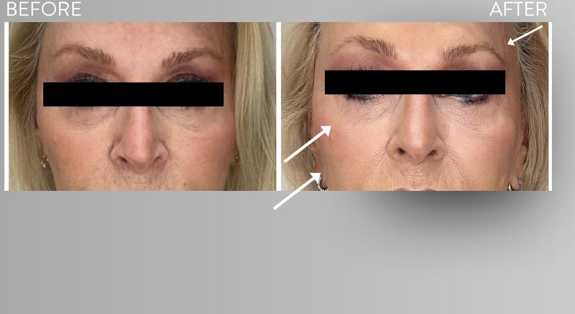
Case 12 · age 76 · 6 treatments
Eye-area comparison
Fuller-looking cheeks and a lighter eye-area appearance. After six sessions, the visible brow arch appears higher, the under-eye area less heavy and the cheek smoother and fuller. These improvements are also apparent in a more mature face. They illustrate the aesthetic direction of muscle conditioning for tissue support; no conclusions are drawn about the obscured eye areas.
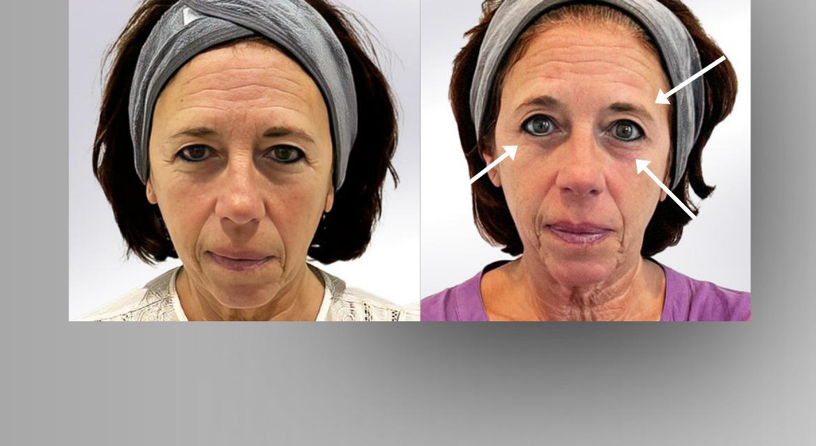
Case 13 · age 49 · 6 treatments
Frontal comparison
Higher-looking brows and a more open gaze. After six sessions, more of the upper eyelid is visible, the brow arch appears higher and under-eye shadow is less prominent. The eye area looks fresher and fine lines softer. This relates to the aim of targeted muscle activation for a more lifted appearance; facial expression is particularly relevant here.
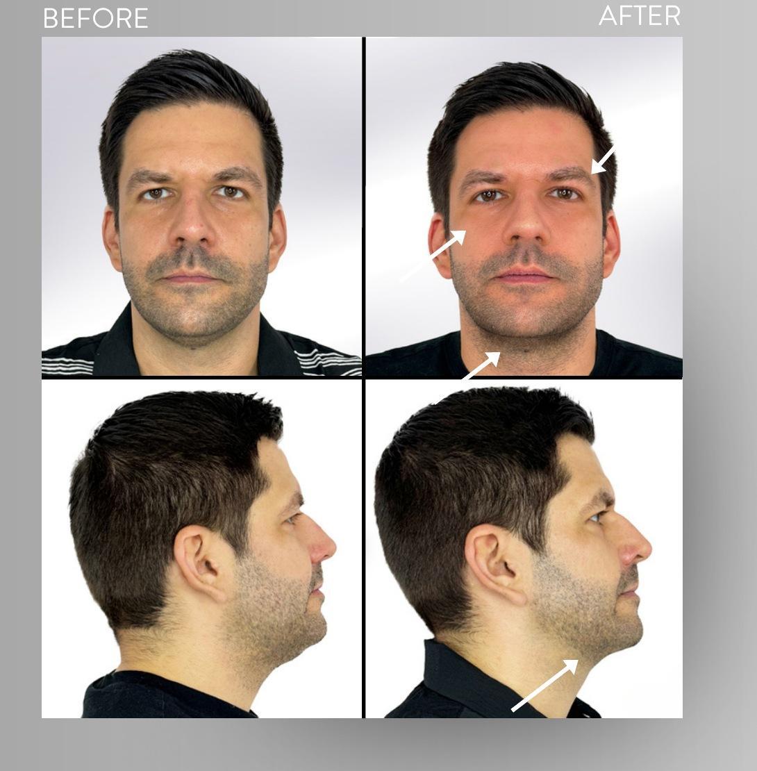
Case 14 · age 40 · 6 treatments
Male face: front and side views
A better-defined profile and fresher eye area. After six sessions, both frontal and profile views show a sharper jawline and a clearer under-chin transition. The eye area looks more open and the cheek firmer. Contour changes visible from several angles illustrate the aesthetic aim of conditioning the lower-face and midface muscles.
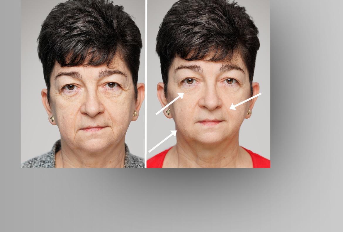
Case 15 · age 62 · 6 treatments
Frontal comparison
Softer shadows and more balanced features. After six sessions, periocular and nasolabial shadows appear less deep. The cheeks look smoother, fuller and more lifted, with a more harmonious mouth area. This refreshed appearance across several regions relates to the tissue-supporting approach of facial muscle conditioning.
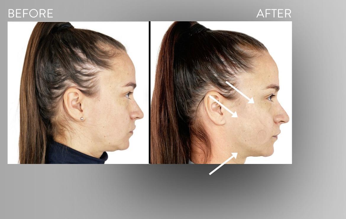
Case 16 · age 38 · 5 treatments
Profile comparison
A lighter lower face and more lifted cheek contour. After five sessions, the profile shows a clearer jaw-to-under-chin transition and less apparent lower-face heaviness. The cheek looks fuller and its surface firmer. This profile change aligns with the contour-supporting aim of deeper facial muscle work; the photograph cannot quantify a change in volume.
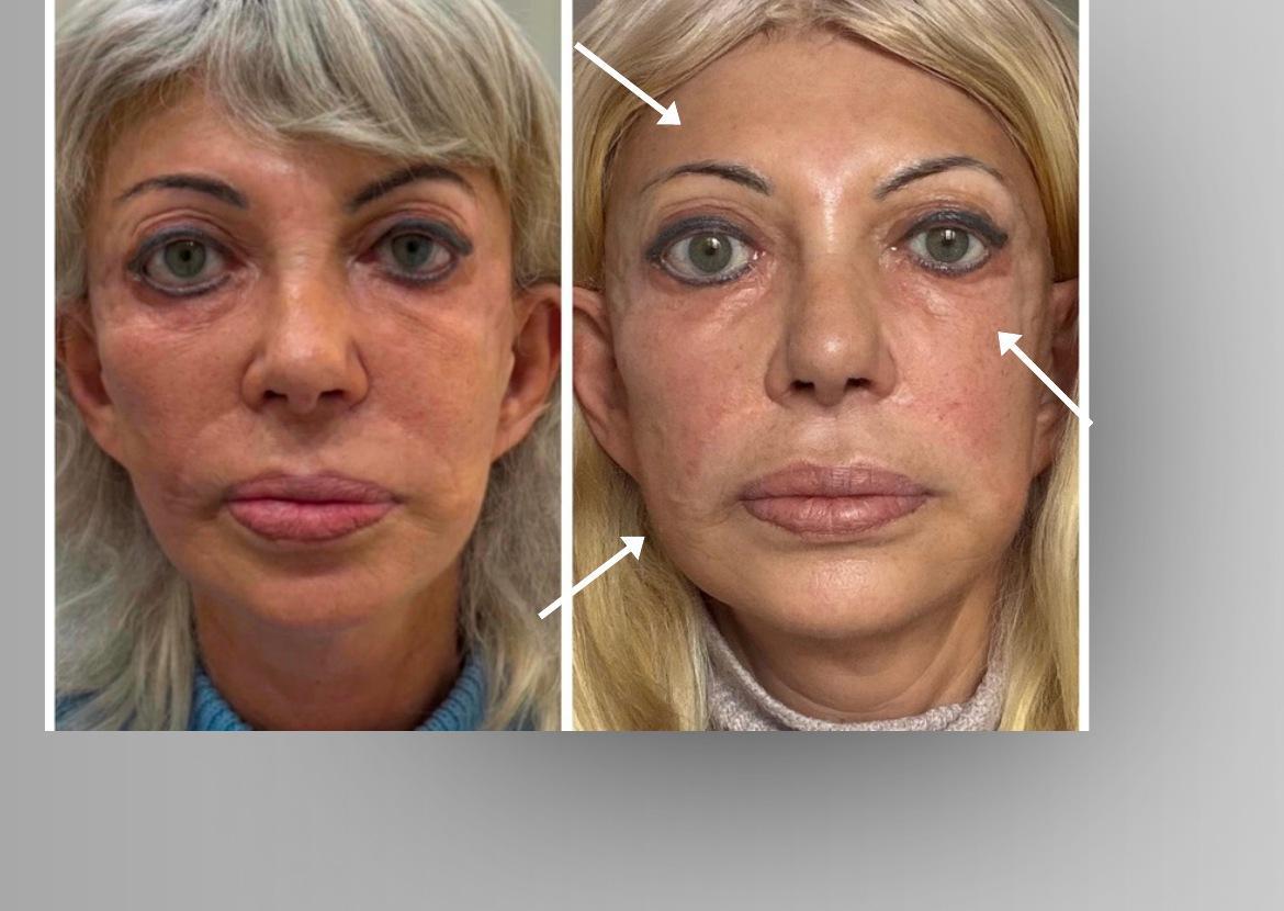
Case 17 · age 61 · 30 treatments
A longer treatment course
A more harmonious contour after an extended course. After thirty sessions, the midface looks fuller and the cheeks better supported; the brow and under-eye areas appear fresher. The surface looks smoother and the facial outline more balanced. This illustrates the long-term approach of regular muscle conditioning, although the photographs do not measure persistence after treatment ends.
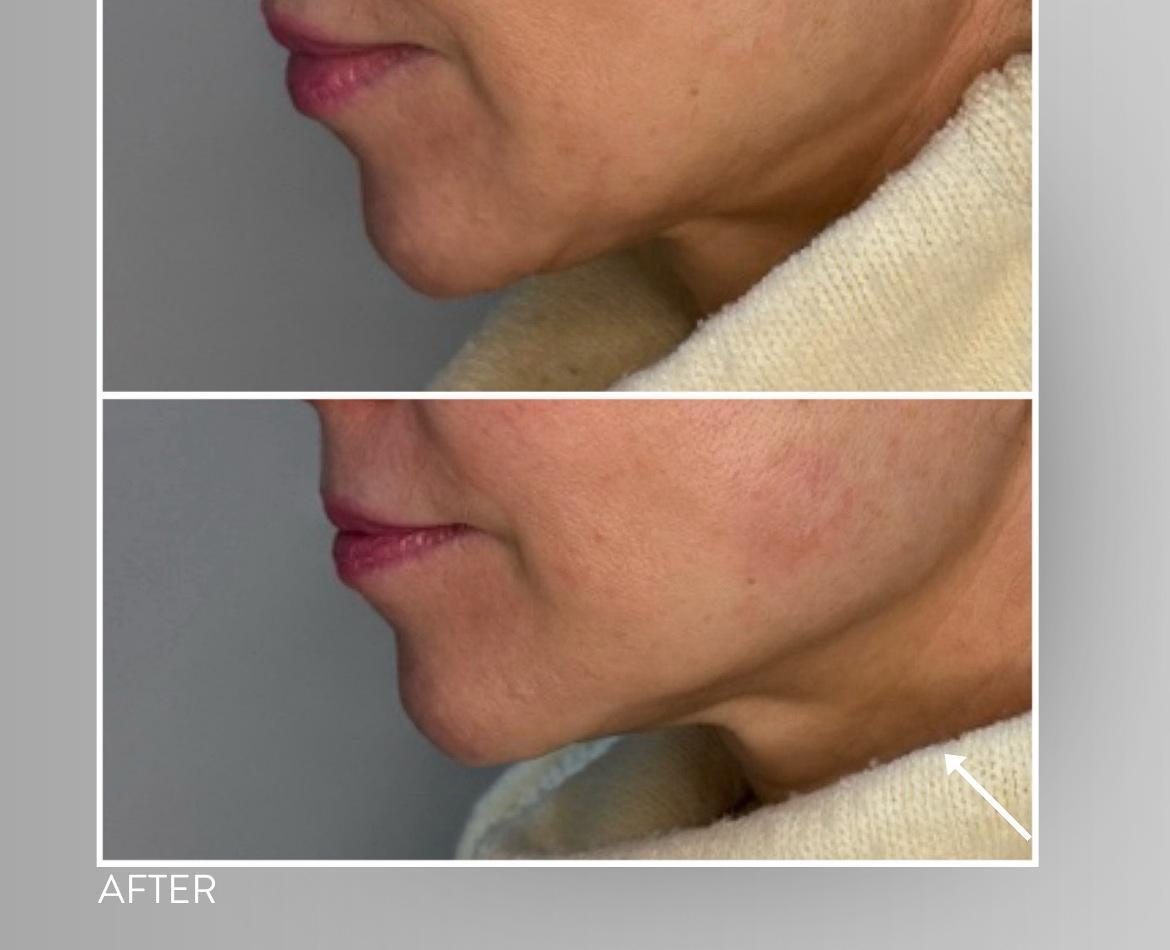
Case 18 · age 49 · 1 treatment
Profile after one treatment
An immediately more defined jawline. The close-up comparison after one session shows a clearer mandibular edge and a more distinct under-chin transition. The lower-face surface looks firmer and the contour more lifted. This is a local, immediate appearance change following muscle activation; it does not establish the lasting muscle conditioning targeted by regular treatment.
18 photographic cases
LifeViz 3D: the facial surface in three dimensions
Three video cases present treatment-associated surface changes: the first uses vector and volume maps, the second shows local displacement measurements from multiple angles, and the third compares the brow and eye area before and after treatment. Below them are four further image-based cases, each with a comparison associated with five treatments. Measurements are case-specific and cannot be combined into a general percentage improvement.
LIFEVIZ 3D · INDIVIDUAL CASE · 46 SECONDS
Contour elevation and fuller-looking cheeks
Silent video with manual playback. Use full-screen mode to examine the measurement markers.
What do the vectors show?
The arrows illustrate the direction of displacement between the pre- and post-treatment facial surfaces. The case assessment supplied with the recording describes approximately 2–3 mm of local displacement in a lifting direction. This refers to the affected regions, rather than a uniform elevation of the entire face.
Fuller contours along the cheeks
The volume map displays positive volume differences of 0.75 mL and 2.23 mL within the selected cheek regions. These are region-specific geometric differences between two surfaces. They are consistent with fuller-looking cheeks, but do not in themselves demonstrate the formation of new muscle tissue.
Targeted muscle activity
ULTIMA engages the facial muscles through repeated, controlled contractions. Treatment aims to support muscle conditioning and soft-tissue support, which may contribute to a firmer, more toned appearance. The surface response shown in the recording may be consistent with this mechanism.
Understanding the immediate response
Active muscle work can increase local metabolic demand and blood flow. Transient changes in tissue fluid distribution and hydration may also contribute to post-treatment fullness. However, 3D imaging does not separately measure muscle-fibre tone, cellular swelling, or the water content of the skin and subcutaneous tissue.
Consistency and follow-up
Longer-term muscle conditioning relies on regularly repeated, individually tailored treatments. A qualified practitioner determines the maintenance schedule. Later follow-up images, acquired under consistent conditions, are needed to distinguish the immediate response from lasting change.
An individual comparison immediately after treatment. The 2–3 mm range is reported in the case assessment and has not been independently remeasured. Image registration, facial expression and the selected measurement region can all influence 3D analysis; individual results vary.
LIFEVIZ 3D · VIDEO CASE 02 · 18 SECONDS
Facial contour changes, point by point
Silent video with manual playback. An excerpt from 00:40–00:58 of the original recording. Use full-screen mode to read the point measurements clearly.
Multiple views, a more detailed comparison
The rotating 3D view allows surface changes in the cheeks, nasolabial region, perioral area and jawline to be followed. The arrows show lateral displacement in some areas and oblique or vertical displacement in others. This enables region-by-region assessment of contour changes within the natural shape of the face.
What do the millimetre values mean?
The excerpt displays local point measurements including 0.57 mm and 1.19 mm. These describe surface differences at the selected points, rather than an average elevation of the whole face. The 2.74 mm marker alongside the colour scale belongs to the display scale; it does not, by itself, establish that amount of lifting. No volume measurement is reported in the selected excerpt.
Supporting facial contours through muscle activity
ULTIMA engages facial muscles through targeted, repeated contractions. The treatment approach focuses on muscle conditioning and support for the natural soft-tissue framework, with the aim of a firmer, more defined facial appearance. Vector imaging provides complementary information by documenting changes at the surface.
Interpreting the surface response
Alongside muscle activity, tissue fluid distribution and facial expression during image capture can influence facial appearance. The 3D video shows differences between two recorded surfaces; muscle-fibre tone, local blood flow and skin hydration require separate assessment.
The role of consistency
Longer-term muscle conditioning relies on regularly repeated, individually tailored treatments. Maintenance sessions and follow-up images taken under consistent conditions work together to track how facial contours change over time.
An individual before-and-after treatment comparison. The excerpt does not specify the number of treatments or the exact timing of the follow-up image. Image registration, facial expression and measurement-point location can influence the result; lasting effects require later follow-up assessment.
LIFEVIZ 3D · VIDEO CASE 03 · 33 SECONDS
A more open gaze, a lighter eyelid sensation
Silent video with click-to-play controls. An excerpt from 01:04–01:37 of the original recording; use full-screen mode to compare the eye area in detail.
A higher brow position and a more open gaze
In the before-and-after comparison, the brow line appears elevated and upper-eyelid hooding is less pronounced. Together, these changes give the eye area a more open, less tired appearance.
Smoother detail around the eyes
Fine lines around the eyes appear smoother, with a more even appearance at the outer corners. The registered views make it easier to compare the brow arch, upper eyelid and surrounding eye area.
The patient's experience
The patient reported a reduced feeling of heavy eyelids after treatment. This feedback on personal comfort complements the aesthetic changes shown in the images.
An individual case combining visual comparison and patient-reported experience. Results may vary between individuals.
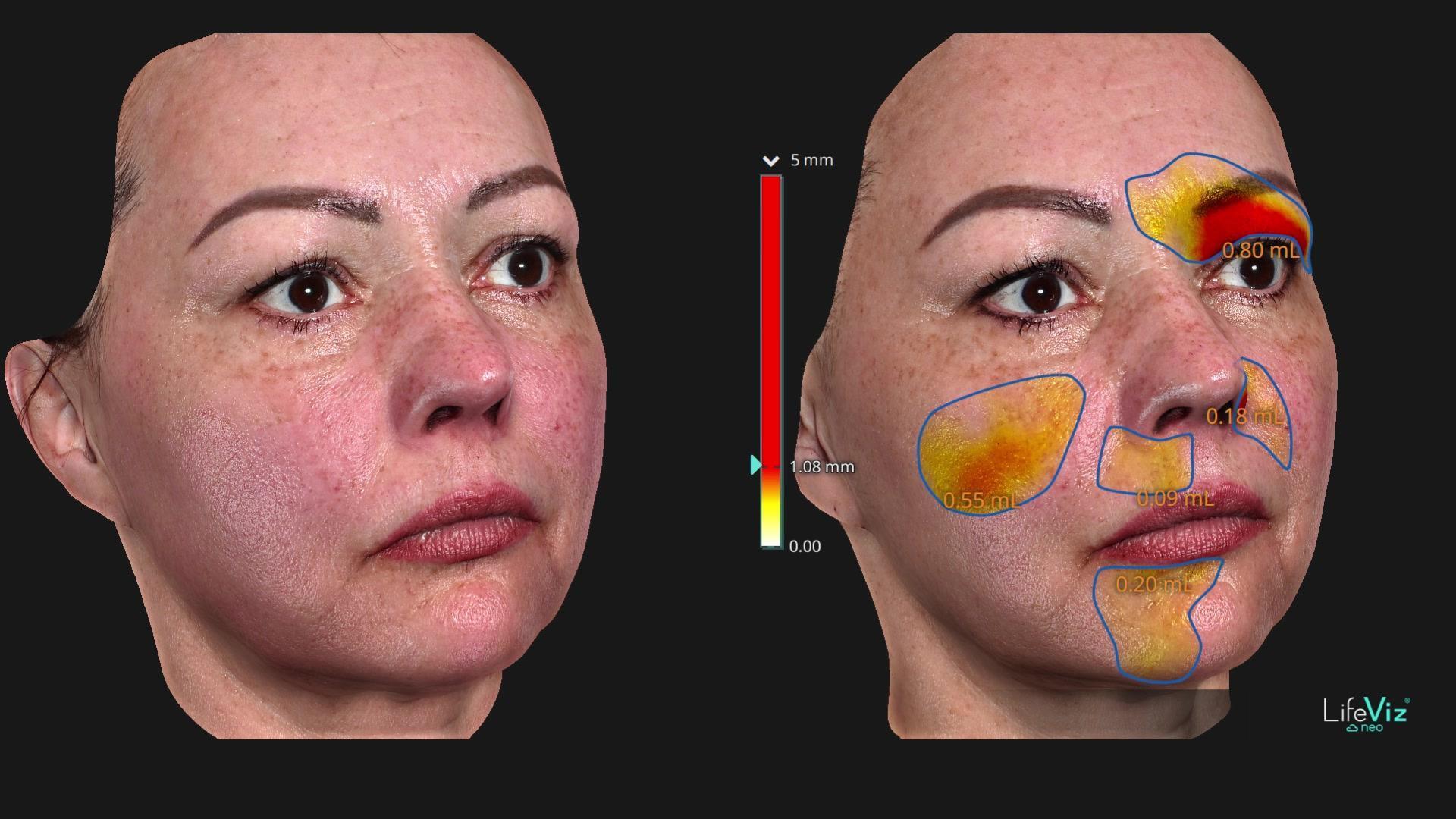
LifeViz 3D · 5 treatments · F–02
F–02 · 3D surface comparison
Local surface differences in the cheek area can be followed. Consistent facial expression also matters when comparing the forehead and lips.
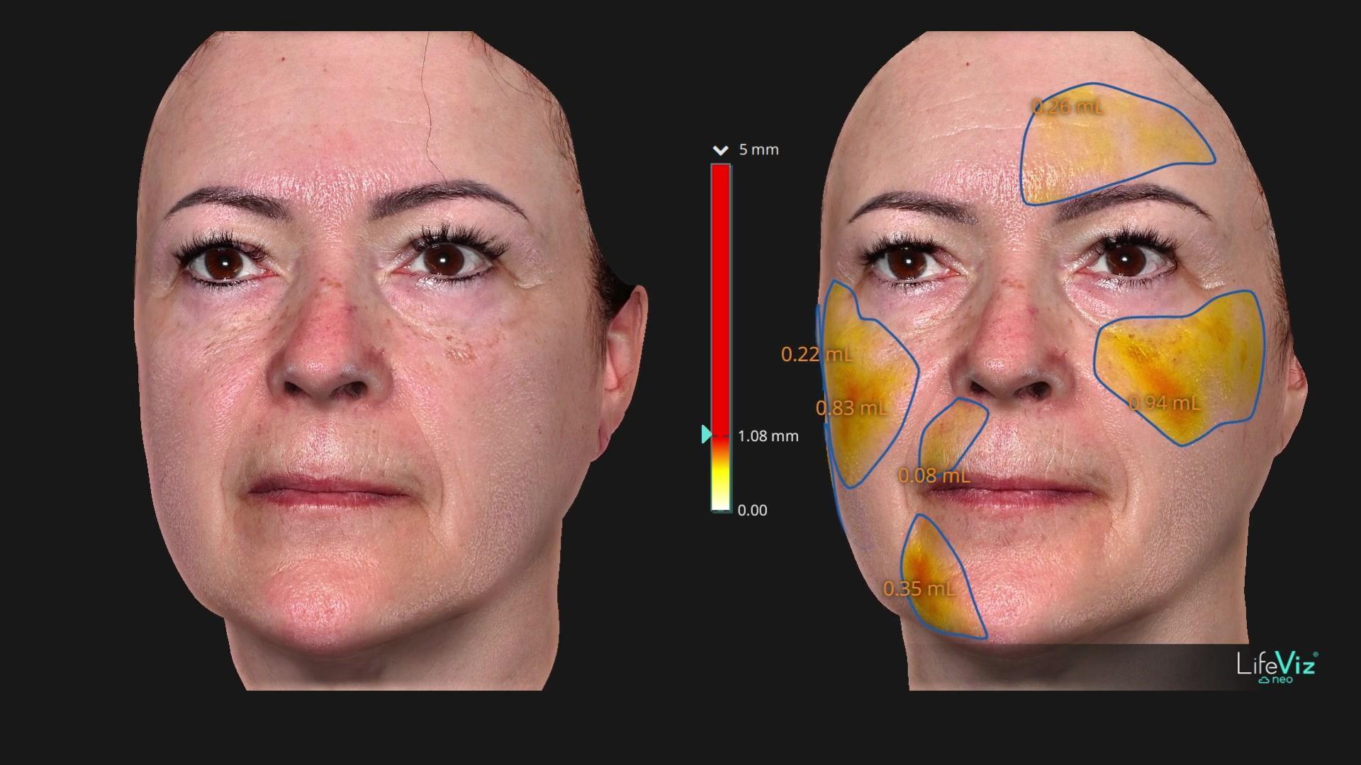
LifeViz 3D · 5 treatments · F–03
F–03 · 3D surface comparison
Within this series, bilateral midface surface changes are most clearly visible here. This compares the geometry of the skin surface.
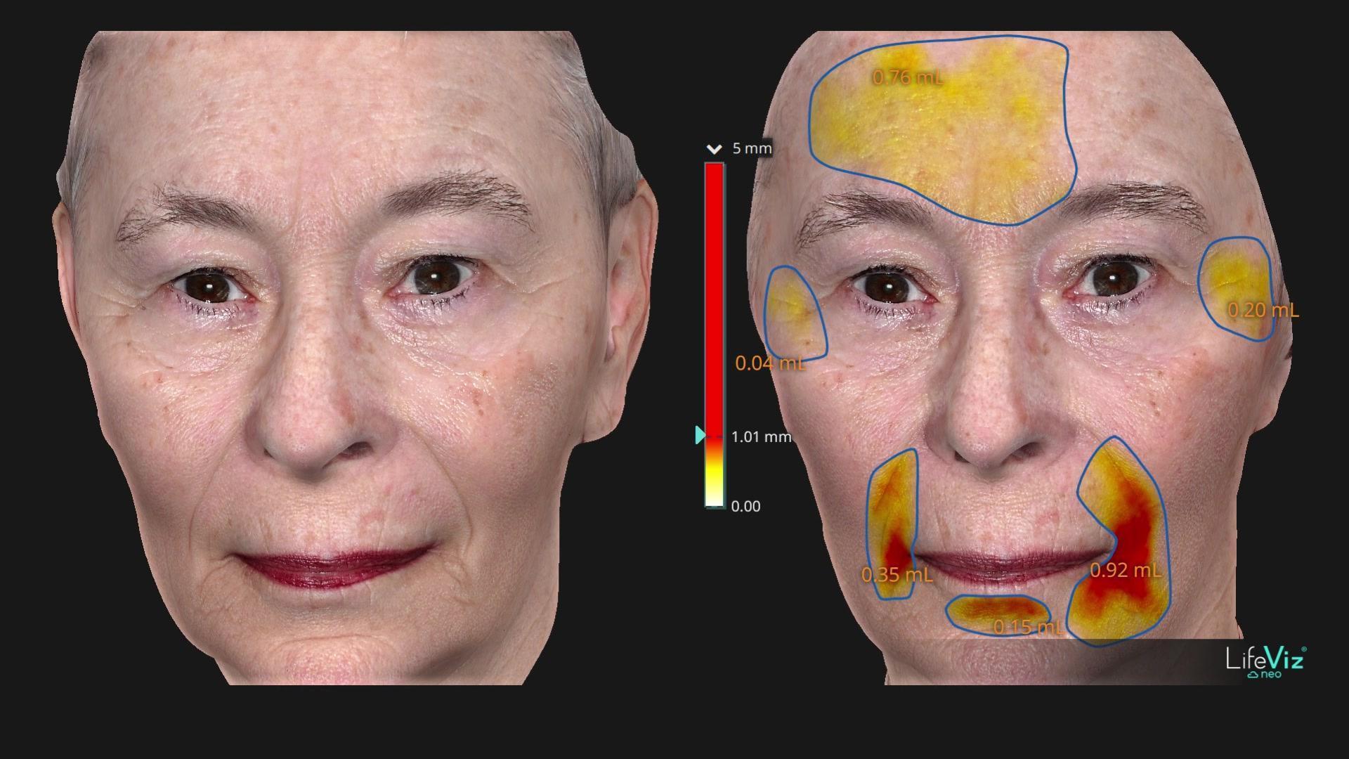
LifeViz 3D · 5 treatments · F–05
F–05 · 3D surface comparison
Lip position also influences comparisons of the perioral areas and the regions beside the nasolabial folds.
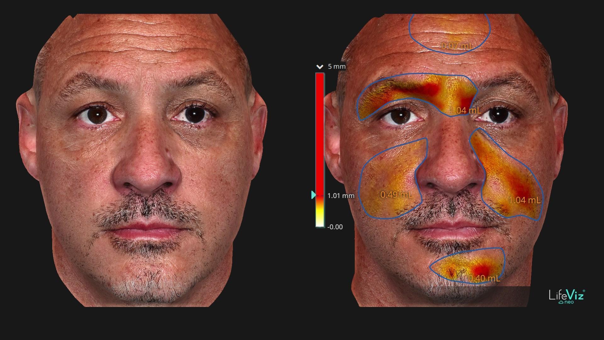
LifeViz 3D · 5 treatments · F–06
F–06 · 3D surface comparison
Both cheek surfaces can be examined together. Differences in eye opening mean the upper face must be assessed cautiously.
Dermus: local ultrasound imaging of the skin
Five original panels with local thickness values in millimetres. The images help interpret the immediate treatment response; on their own, they do not demonstrate new collagen formation or lasting tissue remodelling.
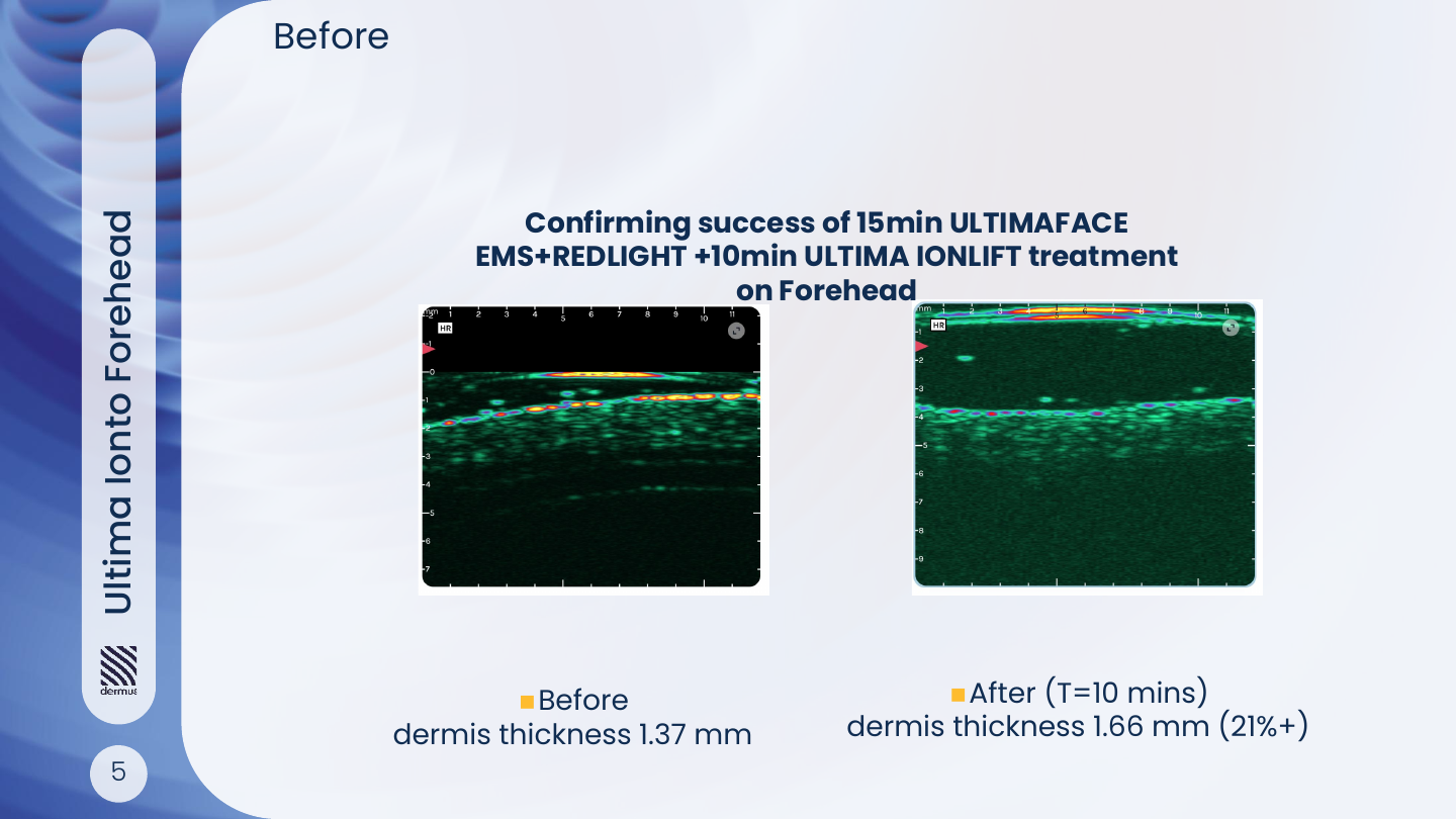
Dermus · local ultrasound measurement
Forehead · FACE + IONLIFT
1.37 → 1.66 mm
The panel shows skin thickness recorded at the identified forehead location before and after the combined treatment.
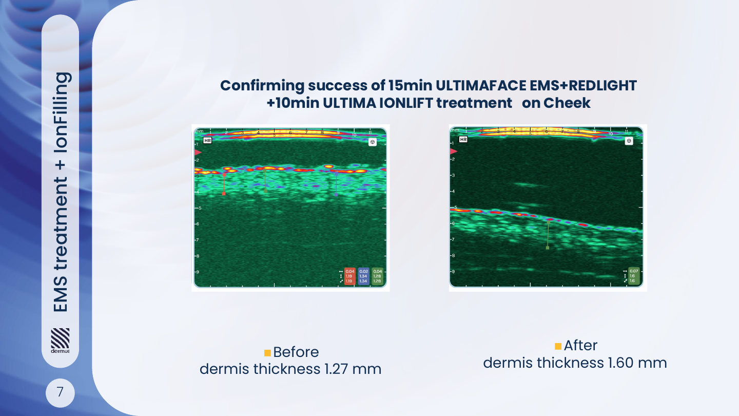
Dermus · local ultrasound measurement
Cheek · FACE + IONLIFT
1.27 → 1.60 mm
The ultrasound comparison of the cheek area illustrates a local measurement associated with the combined treatment.
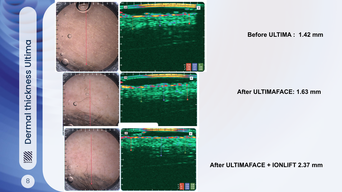
Dermus · local ultrasound measurement
Successive treatment steps
1.42 → 1.63 → 2.37 mm
The three values correspond, in order, to baseline, the measurement after FACE treatment and the measurement after the FACE + IONLIFT combination.
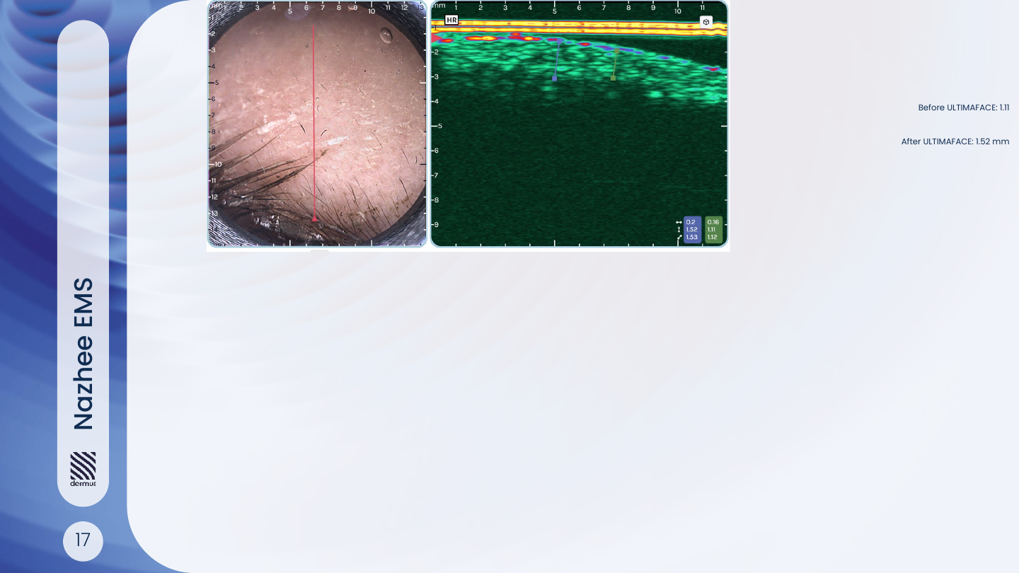
Dermus · local ultrasound measurement
FACE · comparison at two time points
1.11 → 1.52 mm
The paired images show a local thickness measurement before and after FACE treatment.
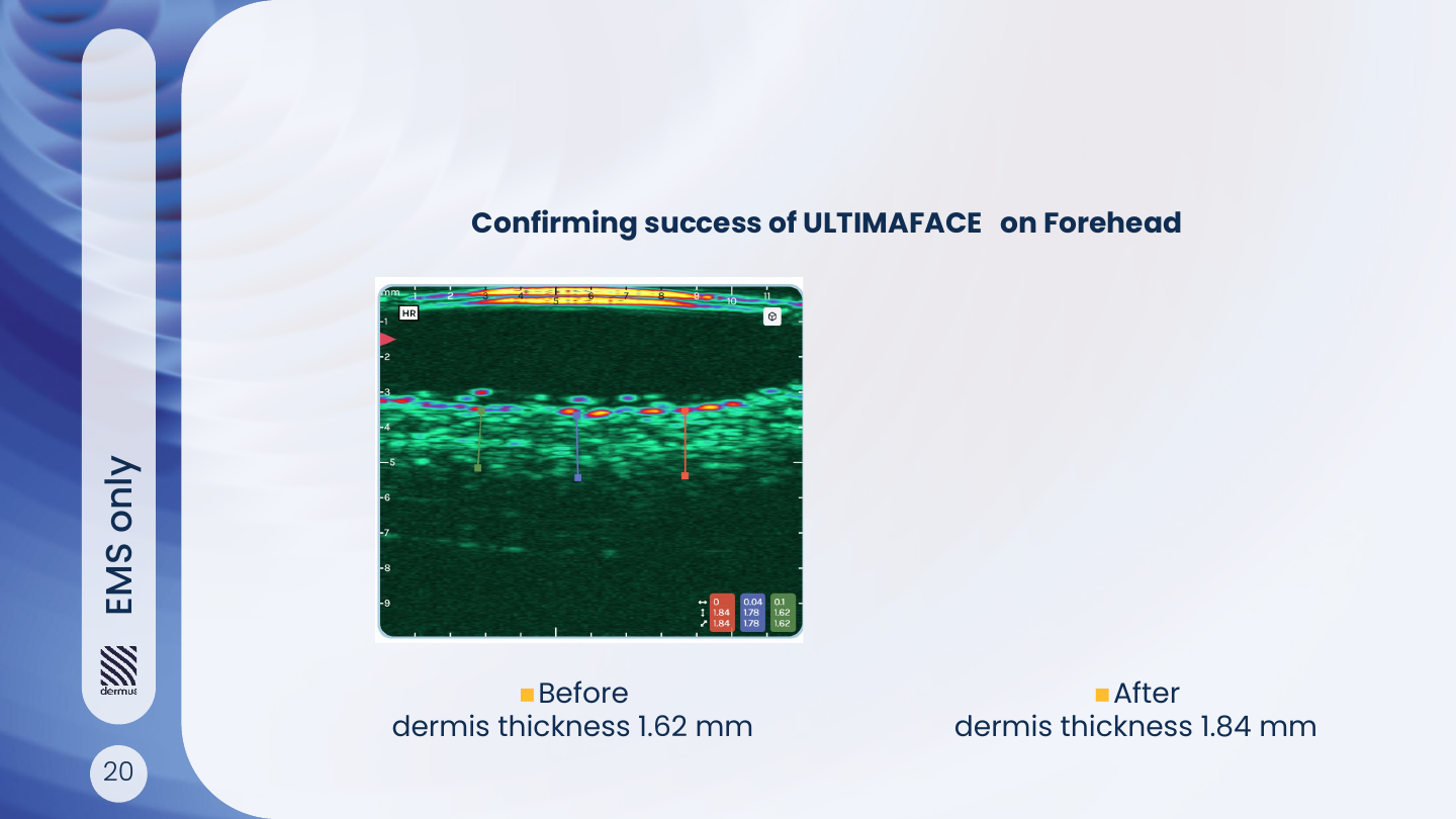
Dermus · local ultrasound measurement
Forehead · muscle stimulation
1.62 → 1.84 mm
The panel compares local forehead ultrasound measurements before and after electrical muscle stimulation.
Featured cases in greater detail
Four selected panels: two photographic and two 3D comparisons. Some cases also appear in the series above. The number of panels therefore does not represent the number of distinct participants.
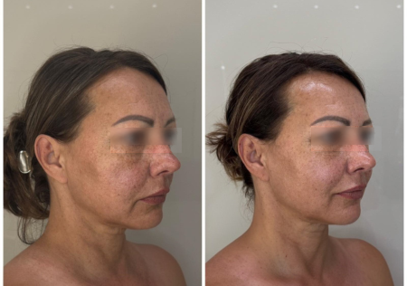
age 54 · 6 treatments
Lower face and midface
This featured comparison illustrates a fuller-looking midface and the contour of the lower face. The same case is also available as Case 01 in the photographic gallery.
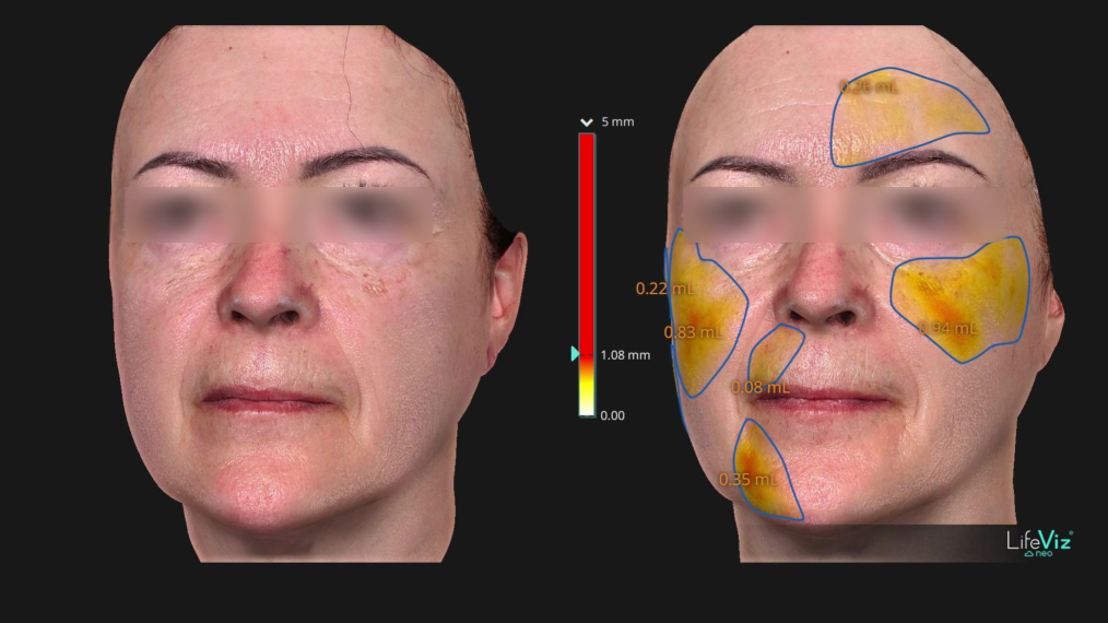
F–03 · LifeViz 3D · 5 treatments
The midface in three dimensions
A featured panel from case F–03, with the eye area concealed. The bilateral midface surface pattern can be followed clearly; the colour map is not a measurement of muscle mass or collagen.
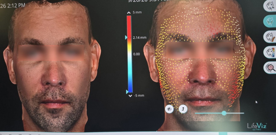
LifeViz 3D · 1 treatment
A single session, an immediate observation
This comparison of a male face illustrates upward surface displacement in parts of the forehead and cheeks. It is an immediate surface response associated with one session, not evidence of a lasting result.
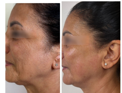
age 56 · 6 treatments
Profile and submental area
The profile images allow comparison of the skin, midface and submental area. The same person appears in Case 05 of the photographic gallery; later follow-up images are also needed to assess persistence of the changes.
How should images be compared?
Always compare before-and-after images of the same person. Lighting, head position, facial expression and imaging time all matter. The datasets presented are observational and may partly overlap; they do not form a single pooled clinical study.
Authorised treatment functions may vary by country.
IMAGES AND CONTEXT
Three methods.
Three types of information.
The different images complement one another. Together they provide a more detailed picture of an individual case, but each records different characteristics.
01
A photograph shows appearance.
Photographs allow comparison of facial contours, proportions, profile and the visible appearance of the skin. Consistent lighting, distance, head position and facial expression support reliable comparison.
A photograph alone does not measure skin thickness, muscle mass or collagen content.
02
A 3D image maps the surface.
LifeViz enables comparison of the shape of the three-dimensional facial surface. Colour maps, displacement markers and selected regions show where and how the two recorded surfaces differ.
Facial expression and body position can also alter the surface. A geometric difference is not the same as a change in the amount of a particular tissue.
03
Ultrasound examines local structure.
The Dermus ultrasound image shows a cross-section of the selected skin area and the thickness between the marked boundaries. Values reported in millimetres relate to the specific location and imaging time point.
Immediately after treatment, fluid distribution, hydration and changes in local circulation may also affect the image. Follow-up measurements under consistent conditions are needed to assess how long these changes last.
FREQUENTLY ASKED QUESTIONS
Behind the visible change,
there is an explanation.
Photographs, three-dimensional facial images and ultrasound examinations complement one another in presenting the results of ULTIMA FACE treatment: from visible contour changes to measurable responses in the tissues beneath the skin.
What do the before-and-after photographs show?
The documented cases repeatedly show fuller, firmer-looking cheeks, a more clearly defined jawline and a more refined lower-face contour. In some images, the eye area appears more open and refreshed, while the nasolabial folds and crow’s feet appear softer. These changes are consistent with targeted stimulation of the facial muscles and improved tissue support provided by those muscles.
How does 3D facial imaging make change measurable?
LifeViz three-dimensional images illustrate spatial changes in the facial surface. In the analysis of six cases involving five treatments each, the most consistent favourable differences appeared in the cheeks and midface. Colour maps show where the surface position and shape changed, allowing the fuller, more defined appearance seen in photographs to be followed in three dimensions.
What do ultrasound images show beneath the skin?
Dermus high-frequency ultrasound provides images of subsurface tissues. In the ULTIMA FACE, IONLIFT and combined treatment series examined, an increase in measured tissue thickness was recorded immediately after treatment. This immediate response may be associated with changes in hydration, local circulation and tissue fluid distribution, and may help explain the fuller, smoother appearance after treatment.
Why is a course of treatment important?
ULTIMA FACE works the facial muscles through repeated, controlled contractions. During a treatment course, this targeted muscle activity may support a firmer appearance and more defined facial contours. The photographic results presented typically relate to 5–8 treatments; the professional adapts treatment count and intensity to individual characteristics.
What results did participants experience?
In the ULTIMA FACE evaluation involving 25 participants, at least noticeable visual improvement was recorded in 88% after five or seven treatments, and all participants reported satisfaction. Together, the photographic, 3D and ultrasound observations support a fuller, firmer and more defined facial appearance.
Results may vary between individuals. The different imaging methods measure different changes; the persistence of the tissue response observed immediately after treatment can be assessed through later follow-up examinations.
DISCOVER THE RELATED TREATMENTS